
Laboratory Medicine ›› 2021, Vol. 36 ›› Issue (1): 1-7.DOI: 10.3969/j.issn.1673-8640.2021.01.001
XU Runhao1, ZOU Chen1, ZHANG Jie2, LI Min2, ZHANG Shulin1( )
)
Received:2019-10-31
Online:2021-01-30
Published:2021-02-05
CLC Number:
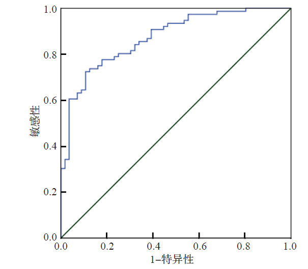
XU Runhao, ZOU Chen, ZHANG Jie, LI Min, ZHANG Shulin. Application of serum bile acid spectrum in the differential diagnosis of pneumonia and lung cancer[J]. Laboratory Medicine, 2021, 36(1): 1-7.
Add to citation manager EndNote|Ris|BibTeX
URL: https://www.shjyyx.com/EN/10.3969/j.issn.1673-8640.2021.01.001
| 组别 | 例数 | TBA/(μmol/L) | CA/(nmol/L) | CDCA/(nmol/L) |
|---|---|---|---|---|
| 肺炎组 | 80 | 4.3(2.9~6.4) | 47.9(24.6~91.8) | 351.5(174.8~718.0) |
| 肺癌组 | 108 | 3.7(2.0~6.2) | 105.7(42.6~369.6)*# | 694.4(312.6~1 823.2)*# |
| 正常对照组 | 106 | 2.7(1.9~4.3) | 46.0(26.5~115.5) | 499.0(188.7~1 018.8) |
| 组别 | DCA/(nmol/L) | LCA/(nmol/L) | UDCA/(nmol/L) | |
| 肺炎组 | 30.9(6.9~108.6)* | 7.9(0.0~15.1)* | 37.2(13.7~111.8)* | |
| 肺癌组 | 246.1(91.9~522.7)# | 15.0(4.8~27.2)# | 123.2(48.8~264.1)# | |
| 正常对照组 | 331.1(161.7~677.6) | 17.1(10.1~29.2) | 150.0(69.6~267.4) | |
| 组别 | 例数 | GCA/(nmol/L) | GCDCA/(nmol/L) | GDCA/(nmol/L) | GLCA/(nmol/L) | GUDCA/(nmol/L) |
|---|---|---|---|---|---|---|
| 肺炎组 | 80 | 256.3 (108.2~473.5) | 948.1 (587.5~1 666.4)* | 33.1 (3.0~132.9)* | 1.6 (0.3~3.3)* | 139.1(72.8~309.1) |
| 肺癌组 | 108 | 229.1 (126.8~464.3) | 868.6 (452.9~1 622.9)* | 160.2 (43.4~373.4)# | 4.1 (1.1~11.7)# | 203.8(87.7~454.6) |
| 正常对照组 | 106 | 165.0 (113.5~300.4) | 621.0 (372.1~1 153.6) | 174.3(74.9~304.7) | 5.4 (1.8~15.6) | 145.8(73.9~358.4) |
| 组别 | TCA/(nmol/L) | TCDCA/(nmol/L) | TDCA/(nmol/L) | TLCA/(nmol/L) | TUDCA/(nmol/L) | |
| 肺炎组 | 25.5(10.9~72.6) | 118.8(50.0~199.9)* | 10.8(2.7~37.0)* | 0.3(0.1~0.8)* | 5.0(1.5~13.6) | |
| 肺癌组 | 21.8(11.3~42.5) | 100.3(53.2~181.9)* | 21.8(8.9~58.3)* | 0.6(0.2~1.3)* | 6.8(3.1~15.1) | |
| 正常对照组 | 21.2(8.6~44.5) | 73.5(40.2~113.6) | 63.7(41.3~90.7) | 1.2(0.4~2.2) | 4.2(2.6~11.1) | |
| 组别 | 例数 | TBA/(μmol/L) | CA/(nmol/L) | CDCA/(nmol/L) |
|---|---|---|---|---|
| 早中期肺癌组 | 53 | 4.0(2.7~6.1) | 89.8(41.4~306.2) | 581.5(305.4~1456.7) |
| 晚期肺癌组 | 51 | 4.2(2.5~6.7) | 186.2(43.5~507.3) | 716.0(476.9~2047.9) |
| Z值 | -1.302 | -1.515 | -1.934 | |
| P值 | 0.188 | 0.130 | 0.066 | |
| 组别 | DCA/(nmol/L) | LCA/(nmol/L) | UDCA/(nmol/L) | |
| 早中期肺癌组 | 237.1(127.4~463.2) | 12.0(7.6~23.5) | 97.3(43.8~249.1) | |
| 晚期肺癌组 | 305.8(152.8~624.0) | 17.9(2.4~29.5) | 146.3(58.3~308.8) | |
| Z值 | -1.810 | -0.328 | -1.310 | |
| P值 | 0.074 | 0.743 | 0.190 | |
| 组别 | 例数 | GCA/(nmol/L) | GCDCA/(nmol/L) | GDCA/(nmol/L) | GLCA/(nmol/L) | GUDCA/(nmol/L) |
|---|---|---|---|---|---|---|
| 早中期肺癌组 | 53 | 261.4(117.0~463.5) | 820.5 (496.0~1 751.9) | 150.7(53.8~311.9) | 4.2 (0.9~7.7) | 189.0(81.4~576.3) |
| 晚期肺癌组 | 51 | 212.1(130.8~479.1) | 977.4 (345.8~1 469.2) | 174.8(52.3~529.1) | 4.3 (1.1~20.6) | 261.5(95.8~416.3) |
| Z值 | -0.335 | -0.257 | -0.824 | -1.083 | -0.166 | |
| P值 | 0.738 | 0.797 | 0.410 | 0.279 | 0.868 | |
| 组别 | TCA/(nmol/L) | TCDCA/(nmol/L) | TDCA/(nmol/L) | TLCA/(nmol/L) | TUDCA/(nmol/L) | |
| 早中期肺癌组 | 24.8(13.4~52.1) | 100.3(64.2~180.8) | 31.8(10.5~62.9) | 0.5(0.2~1.3) | 7.4(3.3~15.0) | |
| 晚期肺癌组 | 18.8(8.9~35.5) | 79.8(41.0~177.0) | 21.1(9.6~47.4) | 0.7(0.2~1.3) | 6.2(2.3~15.3) | |
| Z值 | -1.115 | -1.057 | -0.341 | -0.703 | -0.764 | |
| P值 | 0.265 | 0.291 | 0.733 | 0.482 | 0.445 | |
| 组别 | 例数 | TBA/(μmol/L) | CA/(nmol/L) | CDCA/(nmol/L) |
|---|---|---|---|---|
| 肺鳞癌组 | 34 | 3.4(1.1~6.1) | 92.8(37.9~305.9) | 762.4(232.0~2004.8) |
| 肺腺癌组 | 74 | 3.9(2.2~6.3) | 135.7(42.9~545.1) | 700.9(344.6~1710.5) |
| Z值 | -1.254 | -1.029 | -0.238 | |
| P值 | 0.236 | 0.304 | 0.812 | |
| 组别 | DCA/(nmol/L) | LCA/(nmol/L) | UDCA/(nmol/L) | |
| 肺鳞癌组 | 195.1(114.4~377.7) | 10.4(4.5~22.4) | 113.7(63.7~279.7) | |
| 肺腺癌组 | 265.5(69.0~735.2) | 15.7(6.2~27.4) | 125.0(46.9~240.1) | |
| Z值 | -1.303 | -0.983 | -0.351 | |
| P值 | 0.192 | 0.326 | 0.726 | |
| 组别 | 例数 | GCA/(nmol/L) | GCDCA/(nmol/L) | GDCA/(nmol/L) | GLCA/(nmol/L) | GUDCA/(nmol/L) |
|---|---|---|---|---|---|---|
| 肺鳞癌组 | 34 | 215.1(121.7~377.6) | 868.6 (446.8~1 924.3) | 114.1(58.7~294.3) | 3.2 (1.0~9.6) | 333.5(72.2~641.5) |
| 肺腺癌组 | 74 | 234.6(131.3~481.9) | 926.9 (463.9~1 503.2) | 174.8(41.0~391.9) | 4.3 (1.0~11.8) | 190.7(95.6~415.5) |
| Z值 | -0.377 | -0.930 | -0.902 | -0.298 | -1.005 | |
| P值 | 0.706 | 0.926 | 0.367 | 0.766 | 0.315 | |
| 组别 | TCA/(nmol/L) | TCDCA/(nmol/L) | TDCA/(nmol/L) | TLCA/(nmol/L) | TUDCA/(nmol/L) | |
| 肺鳞癌组 | 22.4(12.2~38.6) | 90.2(54.5~185.1) | 20.2(9.1~62.2) | 0.6(0.3~1.3) | 8.2(3.5~17.3) | |
| 肺腺癌组 | 21.4(10.3~51.3) | 102.7(53.4~182.9) | 25.7(9.1~55.9) | 0.7(0.2~1.3) | 6.3(2.9~12.6) | |
| Z值 | -0.013 | -0.020 | -0.020 | -0.020 | -0.834 | |
| P值 | 0.989 | 0.984 | 0.940 | 0.984 | 0.405 | |
| 组别 | 例数 | CA125/(U/mL) | CA19-9/(U/mL) | CEA/(ng/mL) | CYFRA21-1/(ng/mL) | NSE/(ng/mL) |
|---|---|---|---|---|---|---|
| 肺炎组 | 80 | 16.15(12.55~30.90) | 10.16 (6.91~16.06) | 1.89 (1.34~3.19) | 2.27 (1.73~3.35) | 10.04(9.19~13.10) |
| 肺癌组 | 108 | 19.90(12.84~34.08) | 11.90 (7.28~23.78) | 3.35(1.73~12.23) | 3.29 (1.99~5.51) | 11.15(9.74~14.18) |
| Z值 | -1.122 | -1.621 | -4.546 | -3.337 | -2.430 | |
| P值 | 0.262 | 0.105 | 0.000 | 0.001 | 0.015 |
| 指标 | AUC(95%CI ①) | 最佳临界值 | 敏感性/% | 特异性/% | Youden指数 |
|---|---|---|---|---|---|
| CA | 0.683(0.611~0.749) | 80.55 nmol/L | 59.3 | 73.8 | 0.33 |
| CDCA | 0.670(0.598~0.737) | 574.43 nmol/L | 55.6 | 73.8 | 0.29 |
| DCA | 0.781(0.715~0.838) | 112.46 nmol/L | 73.2 | 77.5 | 0.51 |
| LCA | 0.643(0.570~0.712) | 15.12 nmol/L | 50.1 | 76.3 | 0.26 |
| UDCA | 0.707(0.636~0.771) | 36.04 nmol/L | 86.1 | 50.0 | 0.36 |
| GDCA | 0.657(0.585~0.725) | 57.95 nmol/L | 73.2 | 65.0 | 0.38 |
| GLCA | 0.659(0.587~0.727) | 3.67 nmol/L | 51.9 | 78.8 | 0.31 |
| CEA | 0.704(0.633~0.769) | 2.75 ng/mL | 62.0 | 72.5 | 0.34 |
| CYFRA21-1 | 0.642(0.569~0.711) | 2.36 ng/mL | 69.4 | 57.5 | 0.27 |
| 项目 | B值 | 标准误 | Wald值 | P值 | OR ①(95%CI) |
|---|---|---|---|---|---|
| CA | — | — | 1.136 | 0.287 | — |
| DCA | 0.007 | 0.002 | 16.499 | 0.000 | 1.007(1.004~1.011) |
| CDCA | 0.001 | 0.000 | 8.680 | 0.003 | 1.001(1.000~1.002) |
| LCA | — | — | 0.199 | 0.656 | — |
| UDCA | — | — | 2.782 | 0.095 | — |
| GDCA | — | — | 2.940 | 0.086 | — |
| GLCA | — | — | 1.250 | 0.264 | — |
| CEA | — | — | 2.672 | 0.102 | — |
| CYFRA21-1 | 0.176 | 0.081 | 6.714 | 0.005 | 1.192(1.017~1.398) |
| 常量 | -2.293 | 0.529 | 18.769 | 0.000 | 0.101 |
| [1] | 支修益,石远凯,于金明. 中国原发性肺癌诊疗规范(2015年版)[J]. 中华肿瘤杂志,2015,37(1):67-78. |
| [2] | BRODLIE M,ASEERI A,LORDAN J L,et al.Bile acid aspiration in people with cystic fibrosis before and after lung transplantation[J]. Eur Respir J,2015,46(6):1820-1823. |
| [3] | TOBIN R W,POPE C E 2nd,PELLEGRINI C A,et al. Increased prevalence of gastroesophageal reflux in patients with idiopathic pulmonary fibrosis[J]. Am J Respir Crit Care Med,1998,158(6):1804-1808. |
| [4] | KUIPERS F,BLOKS V W,GROEN A K.Beyond intestinal soap-bile acids in metabolic control[J]. Nat Rev Endocrinol,2014,10(8):488-498. |
| [5] | KHURANA S,RAUFMAN J P,PALLONE T L.Bile acids regulate cardiovascular function[J]. Clin Transl Sci,2011,4(3):210-218. |
| [6] | POLS T W.TGR5 in inflammation and cardiovascular disease[J]. Biochem Soc Trans,2014,42(2):244-249. |
| [7] | ZHANG L,LI T,YU D,et al.FXR protects lung from lipopolysaccharide-induced acute injury[J]. Mol Endocrinol,2012,26(1):27-36. |
| [8] | LIU X,CHEN B,YOU W,et al.The membrane bile acid receptor TGR5 drives cell growth and migration via activation of the JAK2/STAT3 signaling pathway in non-small cell lung cancer[J]. Cancer Lett,2018,412:194-207. |
| [9] | YOU W,CHEN B,LIU X,et al.Farnesoid X receptor,a novel proto-oncogene in non-small cell lung cancer,promotes tumor growth via directly transactivating CCND1[J]. Sci Rep,2017,7(1):591. |
| [10] | 洪庆,周瑛,张有志,等. 我国青年肺癌误诊综述[J]. 临床肺科杂志,2011,16(3):426-427. |
| [11] | I H,CHO J Y. Lung cancer biomarkers[J]. Adv Clin Chem,2015,72:107-170. |
| [12] | 中华医学会放射学分会心胸学组. 低剂量螺旋CT肺癌筛查专家共识[J]. 中华放射学杂志,2015,49(5):328-335. |
| [13] | SUNG H J,CHO J Y.Biomarkers for the lung cancer diagnosis and their advances in proteomics[J]. BMB Rep,2008,41(9):615-625. |
| [14] | JIA W,XIE G,JIA W.Bile acid-microbiota crosstalk in gastrointestinal inflammation and carcinogenesis[J]. Nat Rev Gastroenterol Hepatol,2018,15(2):111-128. |
| [15] | COMEGLIO P,MORELLI A,ADORINI L,et al.Beneficial effects of bile acid receptor agonists in pulmonary disease models[J]. Expert Opin Investig Drugs,2017,26(11):1215-1228. |
| [16] | CHEN B,CAI H R,XUE S,et al.Bile acids induce activation of alveolar epithelial cells and lung fibroblasts through farnesoid X receptor-dependent and independent pathways[J]. Respirology,2016,21(6):1075-1080. |
| [17] | MATSUBARA T,LI F,GONZALEZ F J.FXR signaling in the enterohepatic system[J]. Mol Cell Endocrinol,2013,368(1-2):17-29. |
| [18] | SCHAAP F G,TRAUNER M,JANSEN P L.Bile acid receptors as targets for drug development[J]. Nat Rev Gastroenterol Hepatol,2013,11(1):55-67. |
| [1] | ZHOU Yunlan, SHEN Lisong. Clinical application and challenges of liquid biopsy biomarkers in non-small cell lung cancer [J]. Laboratory Medicine, 2023, 38(9): 807-811. |
| [2] | WANG Huiying, TANG Yu, DONG Lili, WANG Jing. Role of Mycoplasma pneumoniae RNA determination in diagnosis and efficacy monitoring of pneumonia in children [J]. Laboratory Medicine, 2023, 38(9): 865-869. |
| [3] | CHEN Ying, LI Wei, LIN Tao, YANG Li. Relationship between PFTK1 expression level and microvessel density and postoperative prognosis in non-small cell lung cancer [J]. Laboratory Medicine, 2023, 38(7): 634-639. |
| [4] | CHEN Danyang, ZHENG Siyu, ZHENG Ruilin, SU Jingyao, ZHU Bing, LI Yinghua. Research progress in influenza viruses [J]. Laboratory Medicine, 2023, 38(7): 696-703. |
| [5] | ZHU Yurong, ZHANG Dan, HE Yaxing, LI Jingjing, HUANG Jing, LI Ting, LIU Peng, LIU Ronghua. Classification and epidemiology characteristics of Klebsiella pneumoniae isolated from clinic [J]. Laboratory Medicine, 2023, 38(5): 435-440. |
| [6] | YANG Zhongxin, LI Xiaoyu, CHEN Tao, CAO Wenjun. Roles of IL-8 and MMP-2 in the prognosis of non-small cell lung cancer patients after thoracoscopic segmentectomy [J]. Laboratory Medicine, 2023, 38(4): 342-346. |
| [7] | SONG Shiwei, LI Jingwu, LIN Tao, LIU Zeyu, WANG Zhiqiang, SUN Weidong. Relationship between GPNMB and non-small cell lung cancer immune infiltration and prognosis [J]. Laboratory Medicine, 2023, 38(4): 347-351. |
| [8] | WANG Jiawei, ZHU Weinan, CHEN Yingying, JI Ping, WANG Ying. Clinical characteristics,molecular typing and drug resistance genes of carbapenem-resistant Klebsiella pneumoniae isolated from blood culturing [J]. Laboratory Medicine, 2023, 38(4): 362-367. |
| [9] | Expert Group on the Consensus on Androgen Testing in Polycystic Ovary Syndrome China Association for Promotion of Health Science and Technology-Fertility Protection and Preservation Committee. Consensus on androgen testing in polycystic ovary syndrome [J]. Laboratory Medicine, 2023, 38(3): 203-208. |
| [10] | YANG Xiaodong, LI Quanle, PAN Qingqing, ZHOU Jing, XIE Yangmin, ZOU Jihua, SHEN Min, ZHANG Man. Establishment and performance evaluation of candidate reference measurement procedure for the determination of immunosuppressive drugs in human whole blood by ID-LC-MS/MS [J]. Laboratory Medicine, 2023, 38(3): 215-222. |
| [11] | WANG Yaling, HUANG Yunli, TIAN Mi, WANG Shunlan, XIAO Li. Relationship between EOS%,CCR3,eotaxin and disease severity in children with acute bronchopneumonia [J]. Laboratory Medicine, 2023, 38(3): 230-234. |
| [12] | DENG Chen, DAI Jiaze, QIU Lihong, SHEN Weiting, CAI Mufa, LUO Wenying. Molecular epidemic characteristics of hypervirulent Klebsiella pneumoniae in Zhanjiang [J]. Laboratory Medicine, 2023, 38(11): 1026-1031. |
| [13] | HU Shaohua, CHEN Li, ZHAO Meng, MA Zhan, ZHANG Hong. Epidemiological characteristic analysis of Mycoplasma pneumoniae infection in children in Shanghai [J]. Laboratory Medicine, 2023, 38(1): 14-17. |
| [14] | WANG Su, DING Li, JIANG Wenrong, MIAO Yingxin, ZHANG Yanmei, ZHAO Hu. Research progress on drug resistance and treatment strategies of hypervirulent Klebsiella pneumoniae [J]. Laboratory Medicine, 2023, 38(1): 81-86. |
| [15] | LIU Xingqiang, NING Lifen, LI Lin, CHEN Zhongcheng. Correlation of lung cancer clinicopathological characteristics with FR+-CTC,ANXA2 and ProGRP [J]. Laboratory Medicine, 2022, 37(8): 735-740. |
| Viewed | ||||||
|
Full text |
|
|||||
|
Abstract |
|
|||||