检验医学 ›› 2020, Vol. 35 ›› Issue (7): 721-725.DOI: 10.3969/j.issn.1673-8640.2020.07.021

• 技术研究与评价·论著 • 上一篇    下一篇

TRFIA检测胃蛋白酶原在溃疡型胃癌筛查中的价值

杭晨1, 黄飚2, 彭海霞1, 韦欣1, 徐伟红1, 盛慧明1, 李宁丽3   

  1. 1.上海交通大学医学院附属同仁医院,上海 200336
    2.浙江理工大学生命科学与医药学院,浙江 杭州 310018
    3.上海交通大学基础医学院,上海 200025
  • 收稿日期:2019-10-04 出版日期:2020-07-30 发布日期:2020-08-04
  • 作者简介:null

    作者简介:杭 晨,女,1986年生,学士,主管技师,主要从事免疫学检验工作。

  • 基金资助:
    上海交通大学附属同仁医院重点学科资助项目(TR2017xk10)

Role of TRFIA for detecting pepsinogen in screening ulcer gastric cancer

HANG Chen1, HUANG Biao2, PENG Haixia1, WEI Xin1, XU Weihong1, SHENG Huiming1, LI Ningli3   

  1. 1. Tongren Hospital,Shanghai Jiao Tong University School of Medicine,Shanghai 200336,China
    2. College of Life Science and Medicine,Zhejiang Sci-Tech University,Hangzhou 310018,Zhejiang,China
    3. Shanghai Jiao Tong University College of Basic Medical Sciences,Shanghai 200025,China
  • Received:2019-10-04 Online:2020-07-30 Published:2020-08-04

摘要:

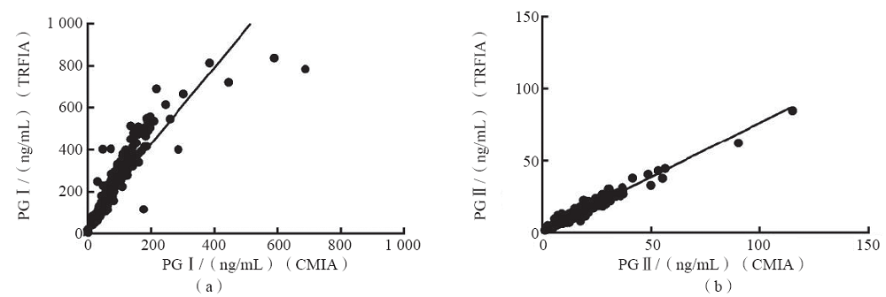
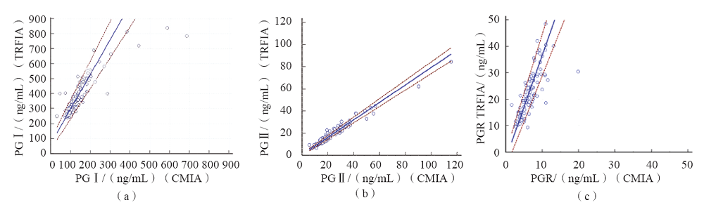
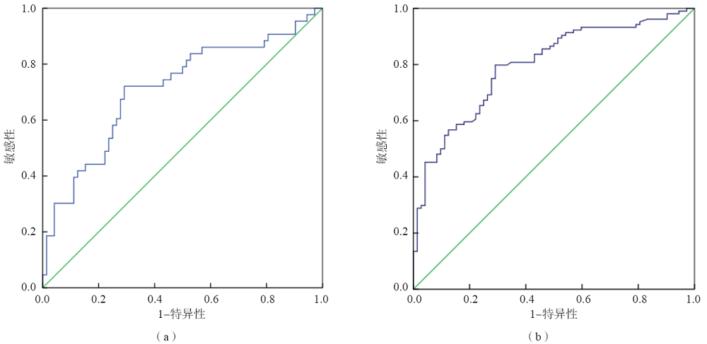
目的 探讨时间分辨荧光分析法(TRFIA)检测血浆胃蛋白酶原(PG)Ⅰ、PGⅡ和胃蛋白酶原Ⅰ/胃蛋白酶原Ⅱ比值(PGR)在溃疡型胃癌筛查中的价值。方法 选取行胃镜检查的患者547例,根据胃镜和病理检查结果分为非萎缩性胃炎72例、非萎缩性胃炎伴有其他病理病变232例、萎缩性胃炎42例、消化性溃疡82例、上皮内瘤变15例、胃癌104例(溃疡型胃癌43例,其他型胃癌61例)。采用TRFIA和化学发光微粒子免疫分析法(CMIA)分别检测其中447名胃病患者血浆PGⅠ、PGⅡ水平,并计算PGR值。采用受试者工作特征(ROC)曲线评价PGR诊断溃疡型胃癌的效能。结果 TRFIA与CMIA检测PGⅠ和PGⅡ的结果均呈正相关(r值分别为0.894、0.982,P<0.05)。TRFIA与CMIA检测PGⅠ高值(TRFIA检测PGⅠ的结果≥240 ng/mL)样本的一致性较差。与消化性溃疡组比较,胃癌组血浆PGⅠ水平及PGR显著降低(P<0.05),溃疡型胃癌组PGR显著降低(P<0.05),其他型胃癌组血浆PGⅠ水平显著降低(P<0.05)。溃疡型胃癌组血浆PGⅡ水平及PGR显著高于其他型胃癌组(P<0.05)。PGR诊断溃疡型胃癌的曲线下面积(AUC)为0.711,最佳临界值为18.20,敏感性为72.1%,特异性为70.8%;诊断胃癌的AUC为0.797,最佳临界值为18.37,敏感性为79.8%,特异性为70.8%。结论 TRFIA与CMIA检测PGⅠ、PGⅡ的结果相关性较好,TRFIA检测上限高于CMIA。PGR对溃疡型胃癌有一定的诊断价值。

关键词: 胃蛋白酶原Ⅰ, 胃蛋白酶原Ⅱ, 胃蛋白酶原Ⅰ/胃蛋白酶原Ⅱ比值, 时间分辨荧光分析法, 溃疡型胃癌, 胃癌

Abstract:

Objective To investigate the role of time-resolved fluoroimmunoassay(TRFIA) for detecting plasma pepsinogen(PG) Ⅰ,PGⅡ and pepsinogen Ⅰ/pepsinogen Ⅱ ratio(PGR) in screening ulcer gastric cancer. Methods According to the results of gastroscopy and pathology,547 patients were classified into 72 cases of non-atrophic gastritis,232 cases of non-atrophic gastritis with other pathology,42 cases of atrophic gastritis,82 cases of peptic ulcer,15 cases of intraepithelial neoplasia and 104 cases of gastric cancer [43 cases of ulcer gastric cancer(ulcer gastric cancer group) and 61 cases of other gastric cancers(other gastric cancer group)]. TRFIA and chemiluminescence microparticle immunoassay(CMIA) were used to detect plasma PGⅠ and PGⅡ levels of 447 patients with gastric diseases,and PGR was calculated. Receiver operating characteristic(ROC) curve was used to evaluate the efficiency of PGR in the diagnosis of ulcer gastric cancer. Results The results of PGⅠ and PGⅡ of TRFIA and CMIA were positively correlated(r=0.894 and 0.982,P<0.05). The results of TRFIA for samples with PGⅠ≥240 ng/mL had poor consistency with CMIA. Compared with peptic ulcer group,plasma PGⅠ level and PGR in gastric cancer group were decreased(P<0.05),and PGR in ulcer gastric cancer group was decreased(P<0.05),and plasma PGⅠ level in other gastric cancer group was decreased(P<0.05). Plasma PGⅡ level and PGR of ulcer gastric cancer group were higher than those of other gastric cancer group(P<0.05). The area under curve(AUC) of PGR for the diagnosis of ulcer gastric cancer was 0.711. The optimal cut-off value was 18.20,the sensitivity was 72.1%,and the specificity was 70.8%. The AUC for the diagnosis of gastric cancer was 0.797,the optimal cut-off value was 18.37,the sensitivity was 79.8%,and the specificity was 70.8%. Conclusions PG Ⅰ and PG Ⅱ detections by TRFIA have a good correlation with CMIA. The upper detection limit of TRFIA is better than that of CMIA. PGR has certain diagnostic value for ulcer gastric cancer.

Key words: Pepsinogen Ⅰ, Pepsinogen Ⅱ, Pepsinogen Ⅰ/pepsinogen Ⅱ ratio, Time-resolved fluoroimmunoassay, Ulcer gastric cancer, Gastric cancer

中图分类号: