检验医学 ›› 2024, Vol. 39 ›› Issue (12): 1173-1180.DOI: 10.3969/j.issn.1673-8640.2024.12.008

• 论著 • 上一篇    下一篇

急、慢性布鲁菌病患者外周血PD-1、LAG-3表达及相关细胞因子水平的变化

朱晓玉1, 李智伟2, 贾金桐1, 李书灵3, 柳叶子2, 王玲玲2, 王倩2, 卢佩佩2, 史清海4()   

  1. 1.石河子大学医学院,新疆 石河子 832000
    2.新疆维吾尔自治区人民医院临床检验中心,新疆 乌鲁木齐 830001
    3.新疆医科大学,新疆 乌鲁木齐 830011
    4.新疆军区总医院全军临床检验诊断中心,新疆 乌鲁木齐 830000
  • 收稿日期:2024-04-21 修回日期:2024-09-19 出版日期:2024-12-30 发布日期:2025-01-06
  • 通讯作者: 史清海,E-mail:shiqinghai@aliyun.com。
  • 作者简介:朱晓玉,女,1997年生,硕士,主要从事布鲁菌病慢性化的免疫机制研究。
  • 基金资助:
    国家自然科学基金项目(81860375);新疆维吾尔自治区自然科学基金面上项目(2023D01C83);新疆维吾尔自治区自然科学基金面上项目(2023D01C84);新疆维吾尔自治区自然科学基金杰出青年科学基金(2022D01E30);新疆维吾尔自治区 “天山英才”培养计划(2022TSYCCX0102);新疆维吾尔自治区科技支疆计划(2022E02118)

Peripheral blood expressions of PD-1,LAG-3 and related cytokines in patients with acute and chronic brucellosis

ZHU Xiaoyu1, LI Zhiwei2, JIA Jintong1, LI Shuling3, LIU Yezi2, WANG Lingling2, WANG Qian2, LU Peipei2, SHI Qinghai4()   

  1. 1. Medical College of Shihezi University,Shihezi 832000,Xinjiang,China
    2. Clinical Laboratory Center,the People's Hospital of Xinjiang Uygur Autonomous Region,Urumqi 830001,Xinjiang,China
    3. Xinjiang Medical University,Urumqi 830011,Xinjiang,China
    4. Clinical Laboratory Diagnostic Center,General Hospital of Xinjiang Military Region,Urumqi 830000,Xinjiang,China
  • Received:2024-04-21 Revised:2024-09-19 Online:2024-12-30 Published:2025-01-06

摘要:

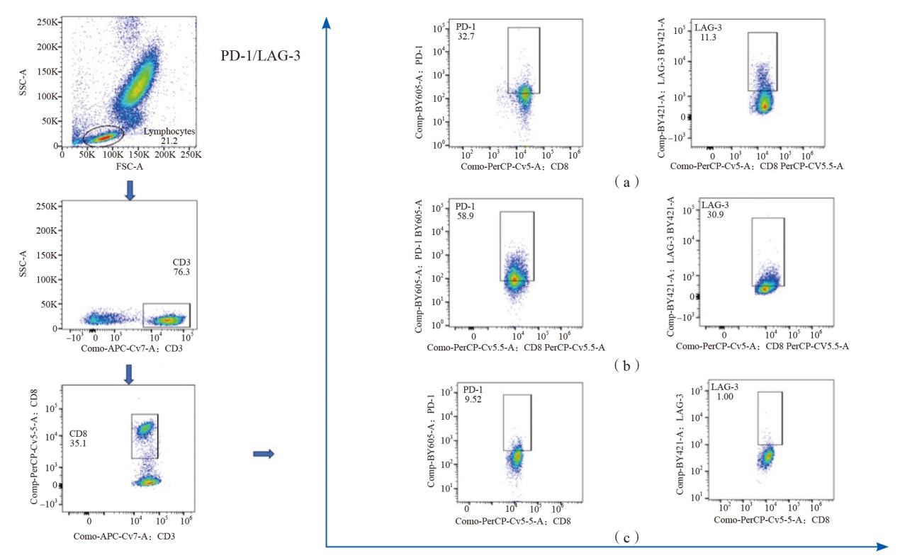
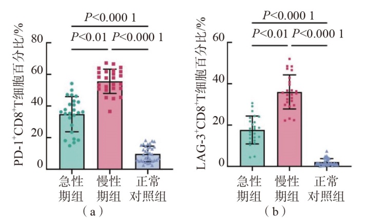
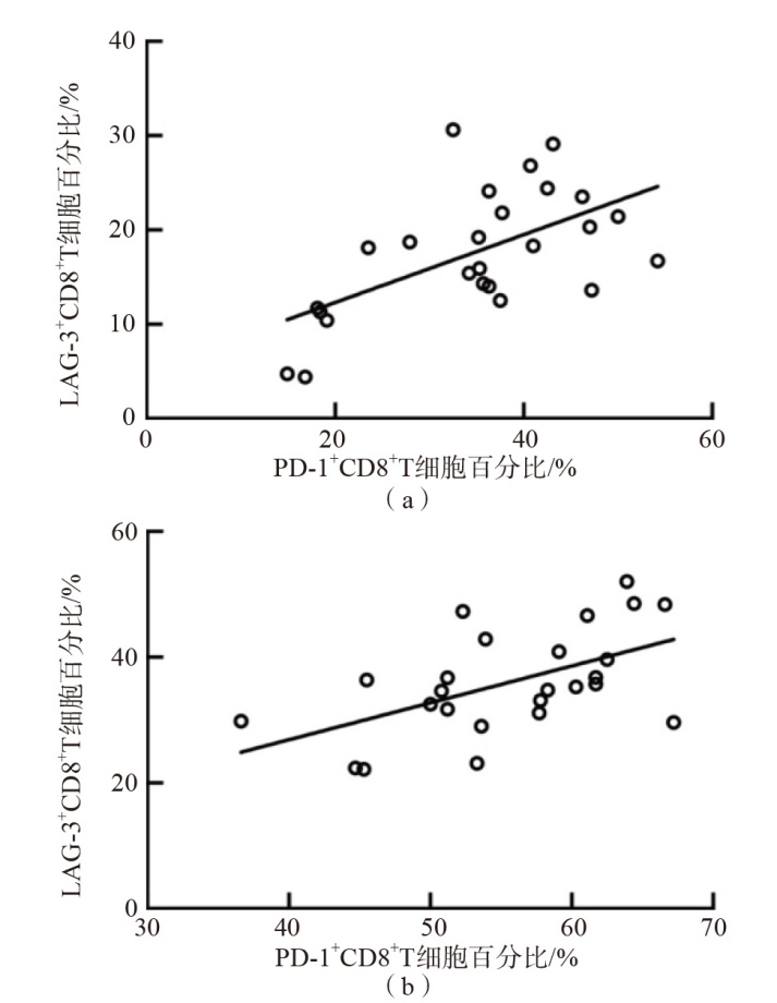
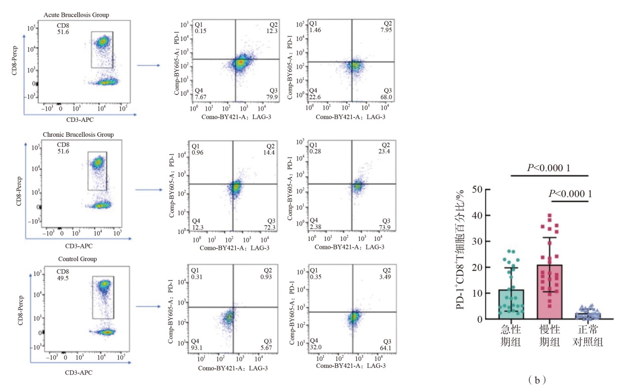
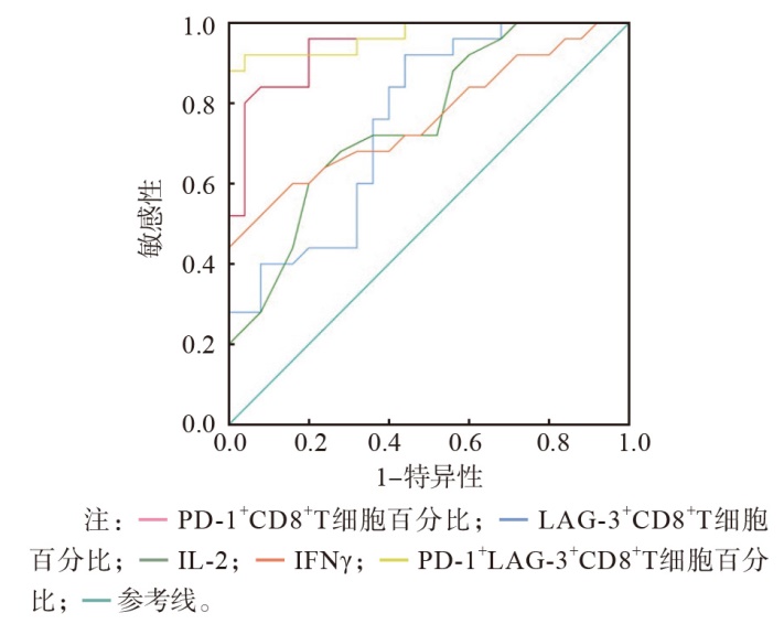
目的 探讨急、慢性布鲁菌病患者外周血CD8+T细胞表面程序性死亡受体1(PD-1)、淋巴细胞活化基因3(LAG-3)表达和血清白细胞介素-2(IL-2)、γ-干扰素(IFN-γ)水平的变化特点。方法 选取2022年12月—2023年8月新疆维吾尔自治区人民医院布鲁菌病患者50例,其中急性期25例(急性期组)、慢性期25例(慢性期组);以同期健康体检者35名作为正常对照组。收集所有研究对象的临床资料和血清学检测结果,并检测外周血CD8+T细胞表面PD-1和LAG-3的表达情况,以及血清IL-2和IFN-γ水平。采用Spearman相关分析评估PD-1和LAG-3表达的相关性。采用受试者工作特征(ROC)曲线评价各项指标区分布鲁菌病急性期和慢性期的效能。结果 与正常对照组比较,急性期组和慢性期组PD-1+CD8+T细胞百分比、LAG-3+CD8+T细胞百分比、PD-1+LAG-3+CD8+T细胞和血清IL-2、IFN-γ水平均显著升高(P<0.000 1)。慢性期组PD-1+CD8+T细胞百分比、LAG-3+CD8+T细胞百分比均高于急性期组(P<0.01),PD-1+LAG-3+CD8+T细胞百分比和血清IL-2、IFN-γ水平2个组之间差异均无统计学意义(P>0.05)。Spearman相关分析结果显示,急性期和慢性期布鲁菌病患者PD-1+ CD8+T细胞百分比与LAG-3+CD8+T细胞百分比呈正相关(rs值分别为0.591、0.545,P<0.01)。PD-1+LAG-3+ CD8+T细胞百分比、LAG-3+CD8+T细胞百分比、PD-1+CD8+T细胞百分比、IFN-γ与病情严重程度均呈正相关(rs值分别为0.726、0.425、0.368、0.395,P<0.05),IL-2与病情严重程度无相关性(rs=0.235,P=0.113)。PD-1+LAG-3+CD8+T细胞百分比、PD-1+CD8+T细胞百分比、LAG-3+CD8+T细胞百分比、IL-2和IFN-γ鉴别布鲁菌病急性期和慢性期的曲线下面积(AUC)分别为0.968、0.945、0.758、0.754、0.762。结论 慢性布鲁菌病患者外周血CD8+T细胞表面抑制性受体PD-1和LAG-3高表达,促炎因子IL-2和IFN-γ下调,慢性布鲁菌病患者可能发生了T细胞耗竭。

关键词: 程序性死亡受体1, 淋巴细胞活化基因3, 白细胞介素-2, γ-干扰素, T细胞耗竭, 布鲁菌病

Abstract:

Objective To study the changes in peripheral blood CD8+T cell surface programmed death receptor 1(PD-1),lymphocyte activation gene 3(LAG-3) expressions and serum interleukin-2(IL-2) and gamma-interferon(IFN-γ) levels in patients with acute and chronic brucellosis. Methods Totally,50 brucellosis patients were enrolled from the People's Hospital of Xinjiang Uygur Autonomous Region from December 2022 to August 2023,of which 25 cases were in acute stage(acute stage group) and 25 cases were in chronic stage(chronic stage group). A total of 35 healthy subjects during the same period were enrolled as healthy control group. The clinical data and serologic determination results of all the patients were collected,and the expressions of PD-1 and LAG-3 on the surface of peripheral blood CD8+T cells,as well as serum IL-2 and IFN-γ levels were determined. Spearman correlation analysis was used to assess the correlation between PD-1 and LAG-3 expressions. The efficacy of each index in acute and chronic stages of brucellosis was evaluated by receiver operating characteristic(ROC) curve. Results Compared with healthy control group,the percentage of PD-1+CD8+T cells,the percentage of LAG-3+CD8+T cells,the percentage of PD-1+LAG-3+CD8+T cells and the levels of serum IL-2 and IFN-γ were higher in acute stage group and chronic stage group(P<0.000 1). The percentage of PD-1+CD8+T cells and the percentage of LAG-3+CD8+T cells in chronic stage were higher than those in acute stage group(P<0.01),and the differences of PD-1+LAG-3+CD8+T cell percentage and serum IL-2 and IFN-γ levels between the 2 groups were not statistically significant(P>0.05). The PD-1+CD8+T cell percentage and LAG-3+CD8+T cell percentage were positively correlated in acute and chronic stage brucellosis patients(rs=0.591 and 0.545,P<0.01). PD-1+LAG-3+CD8+T cell percentage,LAG-3+CD8+T cell percentage,PD-1+CD8+T cell percentage and IFN-γ were positively correlated with disease severity(rs=0.726,0.425,0.368 and 0.395,P<0.05),and IL-2 did not correlate with disease severity(rs=0.235,P=0.113). The areas under curves(AUC) of the percentage of PD-1+LAG-3+CD8+T cells,the percentage of PD-1+CD8+T cells,the percentage of LAG-3+CD8+T cells,IL-2 and IFN-γ in distinguishing acute and chronic stages of brucellosis were 0.968,0.945,0.758,0.754 and 0.762,respectively. Conclusions High expressions of peripheral blood CD8+T cell surface inhibitory receptors(PD-1 and LAG-3) and down-regulation of pro-inflammatory factors(IL-2 and IFN-γ) in patients with chronic brucellosis suggest that patients with chronic brucellosis may undergo T-cell depletion.

Key words: Programmed death receptor 1, Lymphocyte activation gene 3, Interleukin-2, Gamma-interferon, T-cell depletion, Brucellosis

中图分类号: