
Laboratory Medicine ›› 2025, Vol. 40 ›› Issue (3): 223-229.DOI: 10.3969/j.issn.1673-8640.2025.03.004
Previous Articles Next Articles
WANG Shuping, ZHANG Wen, WANG Weiya, MA Lijuan( )
)
Received:2024-11-04
Revised:2025-02-27
Online:2025-03-30
Published:2025-04-10
CLC Number:
WANG Shuping, ZHANG Wen, WANG Weiya, MA Lijuan. Roles peripheral blood lymphocyte subsets and cytokines for viral and autoimmune encephalitis in children[J]. Laboratory Medicine, 2025, 40(3): 223-229.
Add to citation manager EndNote|Ris|BibTeX
URL: https://www.shjyyx.com/EN/10.3969/j.issn.1673-8640.2025.03.004
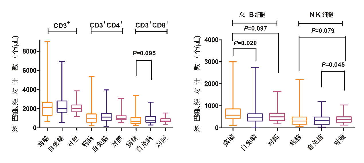
| 组别 | 例数 | CD3+T细胞/ (个·μL-1) | CD3+CD4+T细胞/ (个·μL-1) | CD3+CD8+T细胞/ (个·μL-1) | CD3-CD19+B细胞/ (个·μL-1) | CD3-CD16+CD56+自然杀伤细胞/(个·μL-1) |
|---|---|---|---|---|---|---|
| VE组 | 40 | 2 147.27 (1 321.69,2 668.21) | 1 006.87 (595.50,1 450.24) | 678.90 (468.93,1 077.79)# | 577.96 (438.60,865.26)*# | 307.79 (163.66,494.82)* |
| AE组 | 60 | 2 039.99 (1 642.75,2 812.78) | 1 125.98 (797.41,1 493.55) | 798.02 (604.11,1 165.74) | 456.51 (307.20,635.89) | 336.23 (163.83,491.46)** |
| 正常对照组 | 60 | 2 004.20 (1 668.24,2 384.93) | 1 024.34 (854.31,1 270.17) | 725.37 (657.86,948.86) | 500.75 (328.37,671.58) | 387.81 (262.04,500.47) |
| H值 | 1.559 | 1.746 | 3.037 | 5.449 | 4.970 | |
| P值 | 0.459 | 0.418 | 0.219 | 0.066 | 0.083 |
| 项目 | 例数 | IL-1β/(pg·mL-1) | IL-2/(pg·mL-1) | IL-4/(pg·mL-1) | IL-5/(pg·mL-1) |
|---|---|---|---|---|---|
| VE组 | 40 | 0.34(0.02,9.04)* | 2.22(0.71,4.42)* | 1.83(1.16,2.83)*## | 2.60(1.13,5.65)* |
| AE组 | 60 | 0.28(0.10,2.65) | 1.54(0.38,3.81) | 1.47(1.03,2.17)* | 2.12(0.95,3.17) |
| 正常对照组 | 60 | 0.19(0.03,1.38) | 1.19(0.93,1.68) | 1.19(1.09,1.45) | 1.52(0.92,2.73) |
| H值 | 4.466 | 6.689 | 14.219 | 5.519 | |
| P值 | 0.107 | 0.035 | 0.001 | 0.063 | |
| 项目 | IL-6/(pg·mL-1) | IL-8/(pg·mL-1) | IL-10/(pg·mL-1) | IL-12P70/(pg·mL-1) | |
| VE组 | 3.59(1.39,7.51)* | 4.58(0.34,8.08)*# | 2.34(1.07,2.91)*## | 0.84(0.31,1.71)* | |
| AE组 | 2.30(0.78,5.18)* | 1.51(0.04,4.52)* | 1.25(0.73,2.32)* | 0.52(0.19,1.61) | |
| 正常对照组 | 1.17(0.77,2.05) | 0.62(0.21,1.92) | 0.61(0.45,0.74) | 0.92(0.82,1.03) | |
| H值 | 12.589 | 28.486 | 52.768 | 7.005 | |
| P值 | 0.002 | <0.001 | <0.001 | 0.030 | |
| 项目 | IL-17/(pg·mL-1) | TNF-α/(pg·mL-1) | IFN-α/(pg·mL-1) | IFN-γ/(pg·mL-1) | |
| VE组 | 1.93(0.68,2.75) | 3.26(0.32,6.71)* | 2.79(1.22,6.09)* | 3.60(0.36,9.18)*# | |
| AE组 | 1.78(1.25,2.24) | 2.40(0.58,6.54)* | 2.04(0.53,5.54) | 1.52(0.12,5.76) | |
| 正常对照组 | 1.54(1.19,2.09) | 0.91(0.31,1.63) | 1.75(1.21,2.56) | 1.20(0.52,2.85) | |
| H值 | 1.430 | 7.028 | 5.289 | 4.719 | |
| P值 | 0.489 | 0.030 | 0.071 | 0.094 | |
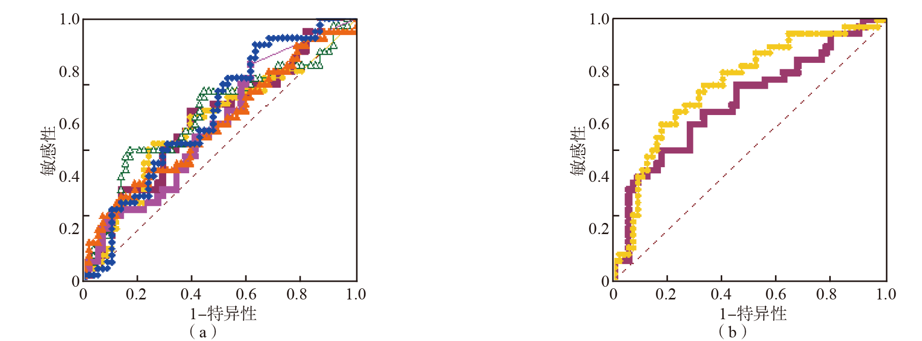
| 项目 | AUC(95%可信区间) | 最佳临界值 | 敏感性/ % | 特异性/ % | 阴性预测值/ % | 阳性预测值/ % | Youden指数 |
|---|---|---|---|---|---|---|---|
| IL-4 | 0.616(0.514~0.712) | >1.63 pg·mL-1 | 65.00 | 60.00 | 72.00 | 52.00 | 0.250 0 |
| IL-8 | 0.603(0.500~0.700) | >4.22 pg·mL-1 | 52.50 | 75.00 | 70.30 | 58.30 | 0.275 0 |
| IL-10 | 0.636(0.534~0.730) | >2.43 pg·mL-1 | 50.00 | 81.67 | 71.00 | 64.50 | 0.316 7 |
| IFN-γ | 0.598(0.496~0.695) | >2.87 pg·mL-1 | 82.50 | 41.86 | 76.70 | 47.10 | 0.243 6 |
| CD3+CD8+T细胞 | 0.599(0.496~0.694) | ≤540.56 个·μL-1 | 32.50 | 86.44 | 65.40 | 61.90 | 0.189 4 |
| CD3-CD19+B细胞 | 0.638(0.535~0.732) | >348.13 个·μL-1 | 90.00 | 37.29 | 84.60 | 48.30 | 0.272 9 |
| 模型A | 0.687(0.587~0.776) | >0.41 | 50.00 | 83.33 | 71.40 | 66.70 | 0.333 3 |
| 模型B | 0.756(0.659~0.836) | >0.67 | 60.00 | 83.05 | 75.40 | 70.60 | 0.430 5 |
| [1] | 中华医学会神经病学分会神经感染性疾病与脑脊液细胞学学组. 中国自身免疫性脑炎诊治专家共识(2022年版)[J]. 中华神经科杂志, 2022, 55(9):931-949. |
| [2] | QIAO S, WU H K, LIU L L, et al. Characteristics and prognosis of autoimmune encephalitis in the east of China:a multi-center study[J]. Front Neurol, 2021, 12:642078. |
| [3] | DUTRA L A, ABRANTES F, TOSO F F, et al. Autoimmune encephalitis:a review of diagnosis and treatment[J]. Arq Neuropsiquiatr, 2018, 76(1):41-49. |
| [4] |
VAISVILAS M, PETROSIAN D, BAGDONAITE L, et al. Seroprevalence of neuronal antibodies in diseases mimicking autoimmune encephalitis[J]. Sci Rep, 2024, 14(1):5352.
DOI PMID |
| [5] | SARAVANOS G L, KING C L, DENG L, et al. Respiratory syncytial virus-associated neurologic complications in children:a systematic review and aggregated case series[J]. J Pediatr, 2021, 239:39-49. |
| [6] | 中国初级卫生保健基金会病原检测专业委员会, 中国医疗保健国际交流促进会分子诊断学分会, 中国研究型医院学会神经科学专委会脑炎协作组. 病毒性脑(膜)炎病原体诊断技术应用专家共识[J]. 中华医学杂志, 2023, 103(9):648-657. |
| [7] | BOCCASAVIA V L, BOVOLENTA E R, VILLANUEVA A, et al. Antigen presentation between T cells drives Th17 polarization under conditions of limiting antigen[J]. Cell Rep, 2021, 34(11):108861. |
| [8] |
ALISSAFI T, KALAFATI L, LAZARI M, et al. Mitochondrial oxidative damage underlies regulatory T cell defects in autoimmunity[J]. Cell Metab, 2020, 32(4):591-604.
DOI PMID |
| [9] | 江载芳, 申昆玲, 沈颖. 诸福棠实用儿科学[M]. 8版. 北京: 人民卫生出版社, 2015. |
| [10] | 关鸿志. 病毒性脑炎的诊治[J]. 中华神经科杂志, 2022, 55(7):747-754. |
| [11] | O'DWYER D N, ASHLEY S L, GURCZYNSKI S J, et al. Lung microbiota contribute to pulmonary inflammation and disease progression in pulmonary fibrosis[J]. Am J Respir Crit Care Med, 2019, 199(9):1127-1138. |
| [12] | BOESEN M S, BORN A P, LYDOLPH M C, et al. Pediatric autoimmune encephalitis in Denmark during 2011-17:a nationwide multicenter population-based cohort study[J]. Eur J Paediatr Neurol, 2019, 23(4):639-652. |
| [13] |
BONNARDEL J, GUILLIAMS M. Developmental control of macrophage function[J]. Curr Opin Immunol, 2018, 50:64-74.
DOI PMID |
| [14] | FRIESER D, PIGNATA A, KHAJAVI L, et al. Tissue-resident CD8+ T cells drive compartmentalized and chronic autoimmune damage against CNS neurons[J]. Sci Transl Med, 2022, 14(640):eabl6157. |
| [15] | 王惠萍, 段丽芬, 孙莹, 等. 自身免疫性脑炎患儿免疫功能与近期预后的相关性分析[J]. 中国免疫学杂志, 2020, 36(23):2899-2903. |
| [16] | SPITERI A G, WISHART C L, PINGET G V, et al. NK cell profiling in West Nile virus encephalitis reveals potential metabolic basis for functional inhibition[J]. Immunol Cell Biol, 2024, 102(4):280-291. |
| [17] | 王蓓红, 张科. 病毒性脑炎患儿脑脊液白介素-6、肿瘤坏死因子、γ干扰素含量测定及其临床意义[J]. 实用临床医药杂志, 2012, 16(17):172-173. |
| [18] |
GARASCHUK O, VERKHRATSKY A. Physiology of microglia[J]. Methods Mol Biol, 2019, 2034:27-40.
DOI PMID |
| [19] | BASU R, HATTON R D, WEAVER C T. The Th17 family:flexibility follows function[J]. Immunol Rev, 2013, 252(1):89-103. |
| [20] | SALIGRAMA N, ZHAO F, SIKORA M J, et al. Opposing T cell responses in experimental autoimmune encephalomyelitis[J]. Nature, 2019, 572(7770):481-487. |
| [21] | PEARL-YAFE M, FABIAN I, HALPERIN D, et al. Interferon-gamma and bacterial lipopolysaccharide act synergistically on human neutrophils enhancing interleukin-8,interleukin-1beta,tumor necrosis factor-alpha,and interleukin-12 p70 secretion and phagocytosis via upregulation of toll-like receptor 4[J]. Shock, 2007, 27(3):226-231. |
| [22] |
ANNUNZIATO F, COSMI L, SANTARLASCI V, et al. Phenotypic and functional features of human Th17 cells[J]. J Exp Med, 2007, 204(8):1849-1861.
DOI PMID |
| Viewed | ||||||
|
Full text |
|
|||||
|
Abstract |
|
|||||