
Laboratory Medicine ›› 2020, Vol. 35 ›› Issue (11): 1177-1185.DOI: 10.3969/j.issn.1673-8640.2020.11.022
Previous Articles Next Articles
GUAN Wenqian, GAO Zhiyuan, FENG Huijuan, HONG Song, HE Yutong, HE Lu, GAO Chunfang
Received:2019-06-17
Online:2020-11-30
Published:2020-12-01
CLC Number:
GUAN Wenqian, GAO Zhiyuan, FENG Huijuan, HONG Song, HE Yutong, HE Lu, GAO Chunfang. Establishment of Lectin-ELISA for the detection of multi-antennary AAG and its preliminary application[J]. Laboratory Medicine, 2020, 35(11): 1177-1185.
Add to citation manager EndNote|Ris|BibTeX
URL: https://www.shjyyx.com/EN/10.3969/j.issn.1673-8640.2020.11.022
| 组别 | 例数 | AST/(U/L) | ALT/(U/L) | GGT/(U/L) | TB/(μmol/L) | |||
|---|---|---|---|---|---|---|---|---|
| HCC组 | 220 | 29.00(20.00~42.00) | 29.00(20.00~42.00) | 56.00(34.00~104.5) | 14.00(10.90~18.38) | |||
| CHB组 | 43 | 223.00(88.00~519.00) | 295.00(183.00~767.00) | 114.00(63.00~165.00) | 23.40(14.70~69.10) | |||
| LC组 | 120 | 32.00(24.25~44.75) | 27.00(18.00~40.25) | 46.50(28.25~79.75) | 23.85(15.00~40.15) | |||
| ICC组 | 74 | 23.00(18.00~35.00) | 24.50(16.00~34.25) | 84.50(61.75~113.00) | 14.35(10.70~18.43) | |||
| 正常对照组 | 80 | 20.00(16.00~25.75) | 14.65(12.00~16.63) | |||||
| 统计值 | 17.295 | 17.744 | 2.271 | 18.798 | ||||
| P值 | <0.001 | <0.001 | 0.132 | <0.001 | ||||
| 组别 | DBil/(μmol/L) | Alb/(g/L) | TP/(g/L) | AFP/(μg/L) | ||||
| HCC组 | 5.50(4.10~7.10) | 42.01±3.74 | 69.62±8.28 | 61.00(4.70~1 210.00) | ||||
| CHB组 | 9.60(5.20~47.00) | 37.69±4.24 | 69.09±6.26 | 8.31(3.71~45.47) | ||||
| LC组 | 10.20(6.00~20.00) | 33.08±10.18 | 63.63±16.81 | 3.60(2.23~8.84) | ||||
| ICC组 | 5.15(3.85~6.80) | 42.26±3.46 | 68.67±5.12 | 2.90(2.00~4.70) | ||||
| 正常对照组 | 47.91±1.97 | 75.87±3.20 | 3.50(2.60~3.50) | |||||
| 统计值 | 22.056 | 46.845 | 161.700 | 82.406 | ||||
| P值 | <0.001 | <0.001 | <0.001 | <0.001 | ||||
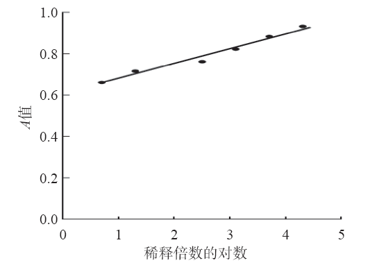
| A值 | x | s | CV/% |
|---|---|---|---|
| 高 | 0.867 | 0.08 | 9.23 |
| 中 | 0.730 | 0.07 | 9.59 |
| 低 | 0.666 | 0.07 | 10.51 |
| A值 | x | s | CV/% |
|---|---|---|---|
| 高 | 0.867 | 0.08 | 9.23 |
| 中 | 0.730 | 0.07 | 9.59 |
| 低 | 0.666 | 0.07 | 10.51 |
| 组别 | DSA-AAG(A值) | AAG/(g/L) |
|---|---|---|
| HCC组 | 0.47±0.07*# | 0.63±0.30* |
| 疾病对照组 | 0.44±0.09* | 0.66±0.36* |
| 正常对照组 | 0.39±0.10 | 0.45±0.15 |
| 组别 | DSA-AAG(A值) | AAG/(g/L) |
|---|---|---|
| HCC组 | 0.47±0.07*# | 0.63±0.30* |
| 疾病对照组 | 0.44±0.09* | 0.66±0.36* |
| 正常对照组 | 0.39±0.10 | 0.45±0.15 |
| 组别 | DSA-AAG(A值) | AAG/(g/L) |
|---|---|---|
| 癌症组 | 0.46±0.08* | 0.67±0.32* |
| 非癌症组 | 0.43±0.10 | 0.52±0.28 |
| 组别 | DSA-AAG(A值) | AAG/(g/L) |
|---|---|---|
| 癌症组 | 0.46±0.08* | 0.67±0.32* |
| 非癌症组 | 0.43±0.10 | 0.52±0.28 |
| 组别 | DSA-AAG(A值) | AAG/(g/L) |
|---|---|---|
| HCC组 | 0.47±0.07* | 0.63±0.30* |
| ICC组 | 0.43±0.10 | 0.79±0.35 |
| 组别 | DSA-AAG(A值) | AAG/(g/L) |
|---|---|---|
| HCC组 | 0.47±0.07* | 0.63±0.30* |
| ICC组 | 0.43±0.10 | 0.79±0.35 |
| 项目 | DSA-AAG(A值) | AAG/(g/L) |
|---|---|---|
| 肿瘤大小/cm | ||
| <5 | 0.47±0.08 | 0.53±0.22 |
| ≥5 | 0.47±0.07 | 0.73±0.33 |
| TNM分期 | ||
| T1期 | 0.47±0.07 | 0.58±0.27 |
| T2期 | 0.47±0.09 | 0.52±0.22 |
| T3+T4期 | 0.47±0.06 | 0.78±0.33 |
| 项目 | DSA-AAG(A值) | AAG/(g/L) |
|---|---|---|
| 肿瘤大小/cm | ||
| <5 | 0.47±0.08 | 0.53±0.22 |
| ≥5 | 0.47±0.07 | 0.73±0.33 |
| TNM分期 | ||
| T1期 | 0.47±0.07 | 0.58±0.27 |
| T2期 | 0.47±0.09 | 0.52±0.22 |
| T3+T4期 | 0.47±0.06 | 0.78±0.33 |
| 组别 | DSA-AAG(A值) | AAG/(g/L) |
|---|---|---|
| AFP阴性HCC组 | 0.48±0.09** | 0.60±0.28* |
| 非癌症组 | 0.43±0.10 | 0.52±0.29 |
| 组别 | DSA-AAG(A值) | AAG/(g/L) |
|---|---|---|
| AFP阴性HCC组 | 0.48±0.09** | 0.60±0.28* |
| 非癌症组 | 0.43±0.10 | 0.52±0.29 |
| 项目 | AUC(95%可信区间) | 最佳临界值 | 敏感性/% | 特异性/% | 准确性/% | 阳性预测值/% | 阴性预测值/% |
|---|---|---|---|---|---|---|---|
| DSA-AAG | 0.651(0.594~0.707) | 0.4 | 85.4 | 42.7 | 63.9 | 58.2 | 77.1 |
| AAG | 0.632(0.577~0.687) | 0.63 g/L | 40.2 | 78.9 | 57.0 | 66.9 | 55.2 |
| AFP | 0.792(0.748~0.836) | 9.80 ng/mL | 66.2 | 84.8 | 74.0 | 77.5 | 71.4 |
| LogitAAG1 | 0.836(0.797~0.874) | 0.52 | 68.5 | 84.2 | 74.9 | 84.6 | 67.0 |
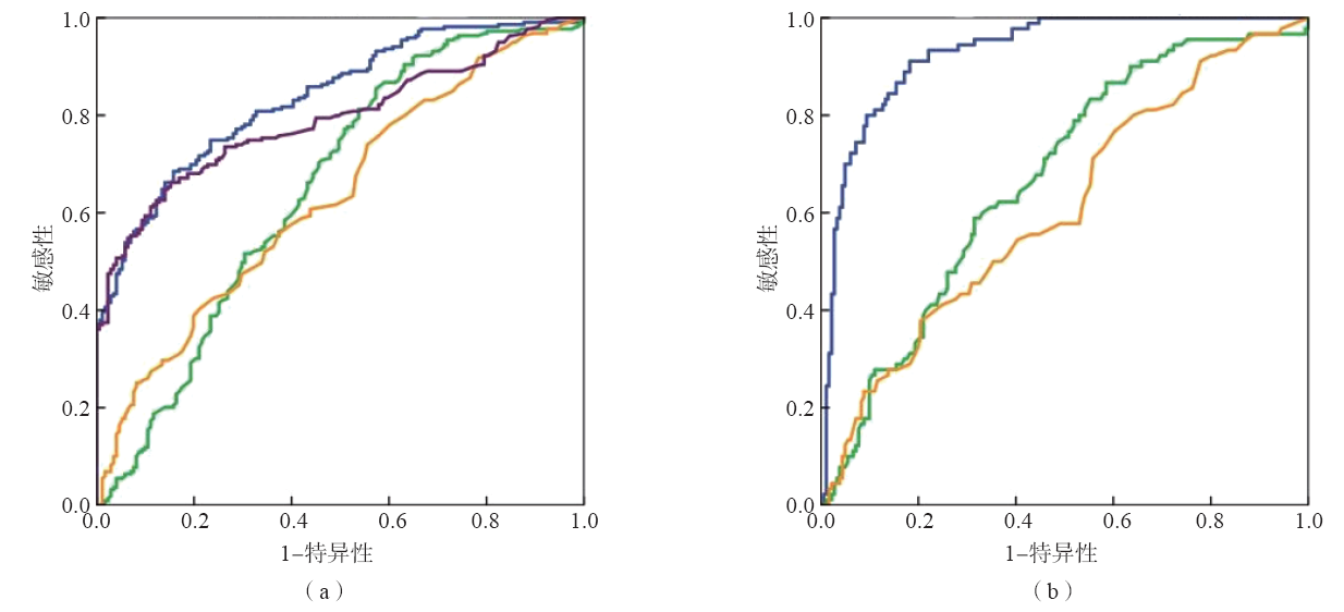
| 项目 | AUC(95%可信区间) | 最佳临界值 | 敏感性/% | 特异性/% | 准确性/% | 阳性预测值/% | 阴性预测值/% |
|---|---|---|---|---|---|---|---|
| DSA-AAG | 0.669(0.603~0.735) | 0.40 | 86.7 | 41.4 | 55.9 | 36.7 | 90.0 |
| AAG | 0.607(0.536~0.678) | 0.64 g/L | 37.8 | 79.6 | 28.4 | 19.3 | 44.4 |
| LogitAAG2 | 0.929(0.899~0.959) | 0.33 | 91.1 | 18.2 | 86.9 | 70.1 | 96.2 |
| [1] | COHN E J,GURD F R N,SURGENOR D M,et al. A system for the separation of the components of human blood:quantitative procedures for the separation of the protein components of human plasma[J]. J Am Chem Soc,1950,72(1):465-474. |
| [2] | KAMIYAMA S,SCHMID K.Studies on the structure of alpha1-acid glycoprotein. Ⅱ. Preparation and characterization of a glycopeptide fraction[J]. Biochim Biophys Acta,1962,58:80-87. |
| [3] | ZHANG C,BI C,CLARKE W,et al.Glycoform analysis of alpha1-acid glycoprotein based on capillary electrophoresis and electrophoretic injection[J]. J Chromatogr A,2017,1523:114-122. |
| [4] | FOURNIER T,MEDJOUBI-N N,PORQUET D.Alpha-1-acid glycoprotein[J]. Biochim Biophys Acta,2000,1482(1-2):157-171. |
| [5] | BERNTSSON J,ÖSTLING G,PERSSON M,et al.Orosomucoid,carotid plaque,and incidence of stroke[J]. Stroke,2016,47(7):1858-1863. |
| [6] | EL-BEBLAWY N M,ANDRAWES N G,ISMAIL E A,et al. Serum and urinary orosomucoid in young patients with type 1 diabetes:a link between inflammation,microvascular complications,and subclinical atherosclerosis[J]. Clin Appl Thromb Hemost,2016,22(8):718-726. |
| [7] | ANDERSEN P,EIKA C.Inhibition of thrombin-induced platelet aggregation by crude and highly purified alpha 1-acid glycoprotein[J]. Scand J Haematol,1980,25(3):202-204. |
| [8] | POS O,OOSTENDORP R A,VAN DER STELT M E,et al. Con A-nonreactive human alpha 1-acid glycoprotein(AGP) is more effective in modulation of lymphocyte proliferation than Con A-reactive AGP serum variants[J]. Inflammation,1990,14(2):133-141. |
| [9] | COSTELLO M,FIEDEL B A,GEWURZ H.Inhibition of platelet aggregation by native and desialised alpha-1 acid glycoprotein[J]. Nature,1979,281(5733):677-678. |
| [10] | RABEHI L,FERRIERE F,SAFFAR L,et al.alpha 1-acid glycoprotein binds human immunodeficiency virus type 1(HIV-1) envelope glycoprotein via N-linked glycans[J]. Glycoconj J,1995,12(1):7-16. |
| [11] | ORCZYK-PAWIĹOWICZ M,KTNIK-PRASTOWSKA I. Terminal monosaccharide expression on amniotic glycoproteins as biomarkers of fetus maturity[J]. Biochem Soc Trans,2011,39(1):344-348. |
| [12] | SAROHA A,BISWAS S,CHATTERJEE B P,et al.Altered glycosylation and expression of plasma alpha-1-acid glycoprotein and haptoglobin in rheumatoid arthritis[J]. J Chromatogr B Analyt Technol Biomed Life Sci,2011,879(20):1839-1843. |
| [13] | TSUTSUMI M,TAKASE S.Usefulness of microheterogeneity of serum alhpa1-acidglycoprotein as a marker for alcohol abuse[J]. Alcohol,2001,25(3):181-184. |
| [14] | MONDAL G,CHATTERJEE U,DAS H R,et al.Enhanced expression of alpha1-acid glycoprotein and fucosylation in hepatitis B patients provides an insight into pathogenesis[J]. Glycoconj J,2009,26(9):1225-1234. |
| [15] | WANG M,ZHU J,LUBMAN D M,et al.Aberrant glycosylation and cancer biomarker discovery:a promising and thorny journey[J]. Clin Chem Lab Med,2019,57(4):407-416. |
| [16] | TANABE K,KITAGAWA K,KOJIMA N,et al.Multifucosylated alpha-1-acid glycoprotein as a novel marker for hepatocellular carcinoma[J]. J Proteome Res,2016,15(9):2935-2944. |
| [17] | TAKETA K,ICHIKAWA E,YAMAMOTO T,et al.Datura stramonium agglutinin-reactive alpha-fetoprotein isoforms in hepatocellular carcinoma and other tumors[J]. Tumour Biol,1990,11(4):220-228. |
| [18] | FANG M,ZHAO Y P,ZHOU F G,et al.N-glycan based models improve diagnostic efficacies in hepatitis B virus-related hepatocellular carcinoma[J]. Int J Cancer,2010,127(1):148-159. |
| [19] | OSNA N A,CARTER W G,GANESAN M,et al.Aberrant post-translational protein modifications in the pathogenesis of alcohol-induced liver injury[J]. World J Gastroenterol,2016,22(27):6192-6200. |
| [20] | OHTSUBO K,MARTH J D.Glycosylation in cellular mechanisms of health and disease[J]. Cell,2006,126(5):855-867. |
| [21] | BLOMME B,VAN STEENKISTE C,GRASSI P,et al.Alterations of serum protein N-glycosylation in two mouse models of chronic liver disease are hepatocyte and not B cell driven[J]. Am J Physiol Gastrointest Liver Physiol,2011,300(5):G833-G842. |
| [22] | CLARK D,MAO L.Cancer biomarker discovery:lectin-based strategies targeting glycoproteins[J]. Dis Markers,2012,33(1):1-10. |
| [23] | 中华人民共和国卫生和计划生育委员会. 原发性肝癌诊疗规范(2017年版)[J]. 传染病信息,2017,30(3):111-127. |
| [24] | WANG M,ZHU J,LUBMAN D M,et al.Aberrant glycosylation and cancer biomarker discovery:a promising and thorny journey[J]. Clin Chem Lab Med,2019,57(4):407-416. |
| [25] | FANG M,ZHAO Y P,ZHOU F G,et al.N-glycan based models improve diagnostic efficacies in hepatitis B virus-related hepatocellular carcinoma[J]. Int J Cancer,2010,127(1):148-159. |
| [26] | ZHANG D,HUANG J,LUO D,et al.Glycosylation change of alpha-1-acid glycoprotein as a serum biomarker for hepatocellular carcinoma and cirrhosis[J]. Biomark Med,2017,11(5):423-430. |
| [27] | MEHTA A,NORTON P,LIANG H Y,et al.Increased levels of tetra-antennary N-linked glycan but not core fucosylation are associated with hepatocellular carcinoma tissue[J]. Cancer Epidemiol Biomarkers Prev,2012,21(6):925-933. |
| [28] | IDE Y,MIYOSHI E,NAKAGAWA T,et al.Aberrant expression of N-acetylglucosaminyltransferase-Ⅳa and Ⅳb(GnT-Ⅳa and b) in pancreatic cancer[J]. Biochem Biophys Res Commun,2006,341(2):478-482. |
| [1] | Clinical Laboratory Society of Chinese Association for Rehabilitation Medicine , Molecular Diagnostics Society of Shanghai Medical Association , Tumor Immunology Branch of Shanghai Society for Immunology , Yueyang Hospital of Integrated Traditional Chinese and Western Medicine of Shanghai University of Traditional Chinese Medicine , Shanghai Center for Clinical Laboratory, Clinical Laboratory Society of Chinese Association of Integrative Medicine , Clinical Laboratory Society of Shanghai Anticancer Association , Tumor Markers Society of Shanghai Anticancer Association . Expert consensus on the clinical application of AFP,AFP-L3% and DCP using GALAD and GALAD-like models in hepatocellular carcinoma [J]. Laboratory Medicine, 2023, 38(7): 607-623. |
| [2] | CHEN Wenju, ZHOU Yong, XU Jiajia, WANG Pan. Role of serum exosomal miR-23b-3p and miR-4429 in patients with hepatocellular carcinoma [J]. Laboratory Medicine, 2023, 38(7): 624-628. |
| [3] | ZHANG Zhixiang, TIAN Hengfeng, HENG Erhu. Genetic test and literature review in a child with congenital glycosylation disorder type Ⅰq [J]. Laboratory Medicine, 2023, 38(2): 137-142. |
| [4] | ZHANG Yifan, LI Changping, ZHANG Lili, CHEN Zhong. Relationship between plasma galectin 3 and low-density lipoprotein cholesterol stratified by ages in patients with cardiovascular disease [J]. Laboratory Medicine, 2023, 38(1): 8-13. |
| [5] | MENG Jun, WANG Junqing, FEI Xiaochun, GU Zhidong. Establishment and validation of a plasma exosome-derived circular RNA model for HCC diagnosis [J]. Laboratory Medicine, 2022, 37(1): 1-10. |
| [6] | TONG Lin, HUANG Chenjun, GAO Zhiyuan, ZHOU Jun, FANG Meng, XIAO Xiao, HE Yutong, HONG Song, XU Minfan, ZHU Feifei, GAO Chunfang. Study on the judgment model of preoperative microvascular invasion in HCC based on common clinical determination items [J]. Laboratory Medicine, 2020, 35(8): 741-748. |
| [7] | JIN Fangfang, JIN Zizheng, LIU Ning, LOU Jinli. Changes of CD3bright T lymphocytes in patients with hepatocellular carcinoma before and after recurrence [J]. Laboratory Medicine, 2020, 35(3): 209-213. |
| [8] | BIE Lihan, FANG Meng, FENG Huijuan, LU Zhicheng, GAO Chunfang. Role of sialylated haptoglobin for the auxiliary diagnosis of hepatitis B-related hepatocellular carcinoma [J]. Laboratory Medicine, 2020, 35(11): 1147-1152. |
| [9] | CHEN Xu, FENG Juan, HU Xiaobo, WANG Changfu. Research progress of pseudouridine as a biomarker of kidney disease and cancer [J]. Laboratory Medicine, 2019, 34(9): 864-867. |
| [10] | MING Xinliang, LIU Xuefang, ZHU Man, TU Jiancheng. Clinical application progress of circulating free nucleic acid in the diagnosis and treatment of hepatocellular carcinoma [J]. Laboratory Medicine, 2019, 34(7): 667-671. |
| [11] | GUAN Wenqian, GAO Chunfang. Research progress of human serum transferrin glycosylation [J]. Laboratory Medicine, 2019, 34(6): 563-566. |
| [12] | ZHENG Xiaorong, WAN Jun, ZHANG Liangying, LI Yongshu, HAN Xuejing, SHI Ping, JIA Kegang. Performance evaluation of galectin-3 detection by chemiluminescence and its preliminary application in heart failure [J]. Laboratory Medicine, 2019, 34(4): 360-366. |
| [13] | FANG Zixiang, ZHANG Xiao, CHEN Yan, WU Qi, ZHU Guoqing, LIU Ya, WANG Jiayi, SUN Fenyong, YU Lei. Role of TFCP2/YAP transcriptional complex co-regulated protein CPE for the diagnosis of hepatocellular carcinoma [J]. Laboratory Medicine, 2019, 34(2): 103-109. |
| [14] | TANG Jie, YANG Yuwei, WANG Kun, YU Jing. Establishment the reference interval of adult protein-induced vitamin K absence/antagonist Ⅱ by chemiluminescent microparticle immunoassay [J]. Laboratory Medicine, 2019, 34(12): 1072-1076. |
| [15] | CAO Minlu, SHEN Minna, ZHOU Yan, WANG Beili, ZHANG Chunyan, PAN Baishen, GUO Wei. Performance comparison of the detection of circulating tumor cells in hepatocellular carcinoma patients by qRT-PCR platform and CellSearch system [J]. Laboratory Medicine, 2019, 34(10): 927-931. |
| Viewed | ||||||
|
Full text |
|
|||||
|
Abstract |
|
|||||