检验医学 ›› 2024, Vol. 39 ›› Issue (2): 138-142.DOI: 10.3969/j.issn.1673-8640.2024.02.007

• 基因组技术与罕见病、遗传病诊治专题 • 上一篇    下一篇

MYH7基因c.1574A>G突变致扩张型心肌病1S型家系分析

路超1, 韩慧娟1, 狄华2, 穆艳超2()   

  1. 1.安阳市中医院心内一科,河南 安阳 455000
    2.安阳市妇幼保健院产前诊断中心 安阳市产前诊断重点实验室,河南 安阳 455000
  • 收稿日期:2023-08-01 修回日期:2023-11-29 出版日期:2024-02-28 发布日期:2024-03-26
  • 通讯作者: 穆艳超
  • 作者简介:穆艳超,E-mail:muyanchao@tmu.edu.cn
    路超,男,1983年生,硕士,副主任中医师,主要从事中西医心血管病分子机制研究。
  • 基金资助:
    安阳市重点研发与推广专项(2023C01SF006);安阳市产前诊断重点实验室项目(2023E02JC003)

Pedigree analysis of dilated cardiomyopathy type 1S caused by MYH7 c.1574AG variation

LU Chao1, HAN Huijuan1, DI Hua2, MU Yanchao2()   

  1. 1. Cardiology Department 1,Anyang Traditional Chinese Medicine Hospital,Anyang 455000,Henan,China
    2. Prenatal Diagnosis Center,Anyang Maternal and Child Health Hospital,Anyang Key Laboratory of Prenatal Diagnosis,Anyang 455000,Henan,China
  • Received:2023-08-01 Revised:2023-11-29 Online:2024-02-28 Published:2024-03-26
  • Contact: MU Yanchao

摘要:

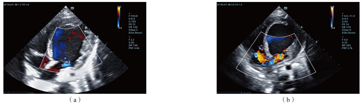
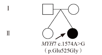
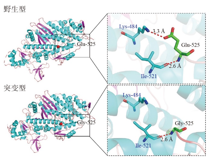
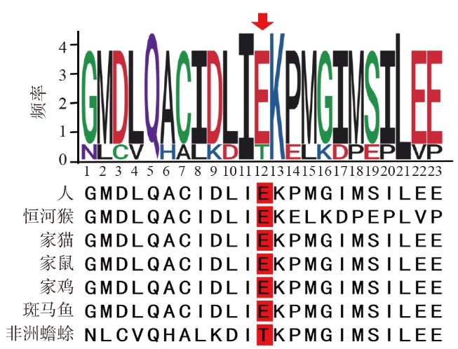
目的 采用全外显子组测序分析1例左心系统扩张患儿的基因变异情况,并进行家系分析,确认扩张型心肌病1S型(DCMIS)的病因。方法 收集1例左心系统扩张患儿的临床资料。采用染色体拷贝数变异测序(CNV-seq)检测患儿染色体结构缺失和重复情况。采用全外显子组测序分析患儿基因变异情况,采用Sanger测序对患儿及其父母、异卵双生姐姐变异位点进行验证。通过生物信息学分析评估变异位点的危害性。结果 患儿心脏彩色多普勒超声示左心功能降低,左心系统明显扩张增大,肺动脉高压,二尖瓣和三尖瓣反流。CNV-seq结果为seq[hg19]46,XN,未发现染色体异常。全外显子组测序分析结果显示,患儿β-心肌肌肉球蛋白重链7(MYH7)基因发生杂合变异[c.1574A>G(p.Glu525Gly)]。检索OMIM、ClinVar数据库,未见相关报道;检索ESP数据库、千人基因组数据库、ExAC数据库和gnomAD数据库,该变异位点未被收录,属于新发变异。Sanger测序证实变异存在,患儿父母、姐姐MYH7基因均正常。MYH7基因c.1574A>G(p.Glu525Gly)变异导致蛋白侧链O端与第484位赖氨酸侧链N端之间形成的氢键侧链相互作用消失。结论 c.1574A>G(p.Glu525Gly)为新发现的MYH7基因变异,是造成患儿左心系统明显扩张的原因。新发变异的检出丰富了DCMIS致病机制研究数据。

关键词: β-心肌肌肉球蛋白重链7基因, 新发突变, 临床特征, 扩张型心肌病1S型

Abstract:

Objective Whole exome sequencing has been used to analyze the variation in one child with left cardiac system dilation,perform pedigree analysis and confirm the etiology of dilated cardiomyopathy type 1S(DCMIS). Methods The clinical data of this child were collected. Chromosome copy number variation sequencing(CNV-seq) was used to determine deletions and duplications in chromosome structure. Whole exome sequencing was also used to analyze the variation in the child,and the variation sites were verified by Sanger sequencing in the child and parents,fraternal sister. The hazard of variation sites was assessed through bioinformatics analysis. Results Color Doppler ultrasound showed decreased left cardiac function,significant dilation and enlargement of left cardiac system,pulmonary hypertension,mitral regurgitation and tricuspid regurgitation. CNV-seq results showed seq[hg19]46,XN,and no chromosome abnormalities were found. Whole exome sequencing showed heterozygous variation in the beta-myosin heavy chain 7(MYH7) gene [c.1574A>G(p.Glu525Gly)]. No relevant reports were found in the OMIM and ClinVar databases. In the ESP database,1000 Genomes database,ExAC database and gnomAD database,the variation site was not included and was a new variation. Sanger sequencing confirmed the existence of variation,and the MYH7 gene of the parents and sister was normal. The MYH7 c.1574A>G(p.Glu525Gly) resulted in the loss of the hydrogen bond side chain interaction between protein side chain O atom and lysine side chain N atom at position 484. Conclusions The c.1574A>G(p.Glu525Gly),a newly discovered variation of MYH7 gene,is responsible for the significant expansion of left cardiac system. The determination of new variation has enriched the understanding of DCMIS pathogenesis.

Key words: Beta-myosin heavy chain 7, New variation, Clinical characteristic, Dilated cardiomyopathy type 1S

中图分类号: