检验医学 ›› 2020, Vol. 35 ›› Issue (12): 1224-1228.DOI: 10.3969/j.issn.1673-8640.2020.12.005

• 外泌体在肿瘤诊疗中的应用专题 • 上一篇    下一篇

血浆外泌体提取方法的比较

吕丽华, 朱丽娜, 王皓, 杨文静, 金安莉, 王蓓丽, 潘柏申, 郭玮()   

  1. 复旦大学附属中山医院检验科,上海 200032
  • 收稿日期:2020-02-29 出版日期:2020-12-30 发布日期:2020-12-31
  • 作者简介:null
    作者简介:吕丽华,女,1992年生,硕士,主要从事外泌体在癌症进展中的作用及机制研究。
  • 基金资助:
    国家自然科学基金面上项目(81772263);国家自然科学基金面上项目(81972000);国家自然科学基金青年基金(81902139);2017年上海市“上海青年临床医技人才(临床检验专业)培养”资助计划;上海市科学技术委员会临床医学领域研究项目(16411952100);上海市临床重点专科建设项目(医学检验科);复旦大学附属中山医院院内临床研究项目(2018ZSLC05)

Comparison of plasma exosome extraction methods

LÜ Lihua, ZHU Lina, WANG Hao, YANG Wenjing, JIN Anli, WANG Beili, PAN Baishen, GUO Wei()   

  1. Department of Clinical Laboratory,Zhongshan Hospital,Fudan University,Shanghai 200032,China
  • Received:2020-02-29 Online:2020-12-30 Published:2020-12-31

摘要:

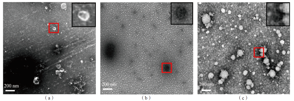
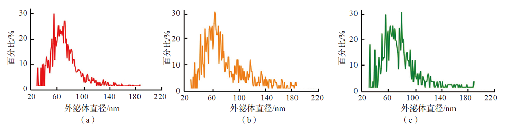
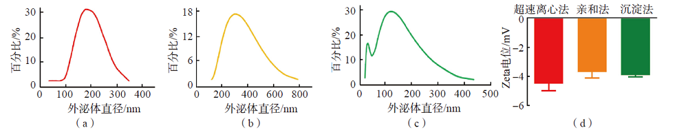
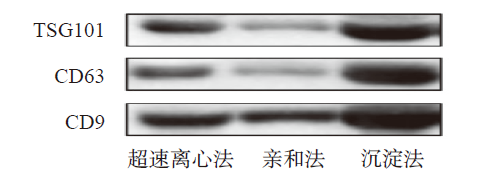
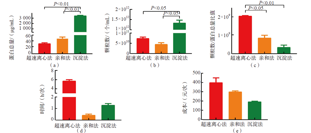
目的 比较超速离心法、膜亲和法、沉淀法提取血浆外泌体的效率。方法 分别采用超速离心法、膜亲和法、沉淀法提取血浆外泌体。采用透射电子显微镜、纳米流式检测仪和动态光散射法检测外泌体的形态、颗粒数、粒径分布及Zeta电位,采用免疫印迹法鉴定外泌体标志性蛋白的表达,采用二喹啉甲酸(BCA)法测定外泌体蛋白浓度,分析颗粒数/蛋白总量比值以评估外泌体的提取纯度。 结果 超速离心法、沉淀法和膜亲和法提取的外泌体在透射电子显微镜下均能观察到其立体膜结构,大小为40~100 nm,但沉淀法提取的外泌体混有聚乙二醇(PEG)聚合物及蛋白大颗粒。亲和法提取的外泌体直径大于超速离心法和沉淀法(P<0.05)。动态光散射法结果显示3种方法提取的外泌体的Zeta电位均为负值,但测得的外泌体直径偏大。免疫印迹法结果显示3种方法提取的外泌体均表达标志性蛋白。沉淀法提取的血浆外泌体蛋白总量和外泌体颗粒数均高于超速离心法和膜亲和法(P<0.05)。超速离心法的提取纯度高于膜亲和法和沉淀法(P<0.05)。结论 超速离心法、膜亲和法和沉淀法均能提取出血浆外泌体,应根据实验需求选择合适的方法。

关键词: 外泌体, 超速离心法, 膜亲和法, 沉淀法

Abstract:

Objective To compare the efficiency of ultracentrifugation,affinity membrane method and precipitation for extracting plasma exosome. Methods Plasma exosomes were extracted by 3 different methods,including ultracentrifugation,affinity membrane method and precipitation. Transmission electron microscopy,nano-flow cytometry,zetasizer nano were used to verify its morphology,particle size and distribution and Zeta potential. The western blot analysis was performed to measure the representative protein markers of exosomes. The bicinchoninic acid assay was utilized to measure concentration of exosomal protein. The ratio of particles number to protein concentration was analyzed to evaluate the extraction efficiency of exosomes. Results The transmission electron microscope showed exosomes extracted by ultracentrifugation and affinity membrane method as a saucer-like bilayer membrane structure,while the exosomes extracted by precipitation were mixed with PEG polymer and protein particles. The size of the exosomes were between 40-100 nm,while the mean diameter of the exosomes derived using affinity membrane method was larger than those using the ultracentrifugation and precipitation. Dynamic light scattering(DLS) analysis showed that the Zeta potential of exosomes was negative,which implied their membranous structure. However,exosomes size measured by DLS were larger than that by nano-flow cytometry. In addition,all the exosomes expressed representative protein markers. The precipitation produced the highest protein concentration and particles,and the ultracentrifugation gave exosomes the highest purity than the other 2 methods. Conclusions Exosomes can be extracted by ultracentrifugation,affinity membrane and precipitation. However,which extraction method should be used depends on the specific experimental requirements.

Key words: Exosomes, Ultracentrifugation, Affinity membrane method, Precipitation

中图分类号: