检验医学 ›› 2024, Vol. 39 ›› Issue (6): 542-547.DOI: 10.3969/j.issn.1673-8640.2024.06.005

• 论著 • 上一篇    下一篇

血管性帕金森综合征患者线粒体自噬信号通路PINK1/Parkin表达及其与神经功能的关系

娄展1, 彭涛2, 刘星亮1, 岳秉宏1, 李燃1, 智永怡1   

  1. 1.河北北方学院附属第一医院神经内科,河北 张家口 075000
    2.河北北方学院附属第一医院内镜中心,河北 张家口 075000
  • 收稿日期:2022-08-04 修回日期:2023-11-13 出版日期:2024-06-30 发布日期:2024-07-08
  • 作者简介:娄 展,女,1987年生,硕士,主治医师,主要从事帕金森综合征等神经系统疾病的诊治工作。
  • 基金资助:
    河北省医学科学研究课题(20200519)

Expression of mitochondrial autophagy signaling pathway PINK1/Parkin and its relationship with neural function in patients with vascular Parkinsonism

LOU Zhan1, PENG Tao2, LIU Xingliang1, YUE Binghong1, LI Ran1, ZHI Yongyi1   

  1. 1. Department of Neurology,the First Affiliated Hospital of Hebei North University,Zhangjiakou 075000,Hebei,China
    2. Endoscopy Center,the First Affiliated Hospital of Hebei North University,Zhangjiakou 075000,Hebei,China
  • Received:2022-08-04 Revised:2023-11-13 Online:2024-06-30 Published:2024-07-08

摘要:

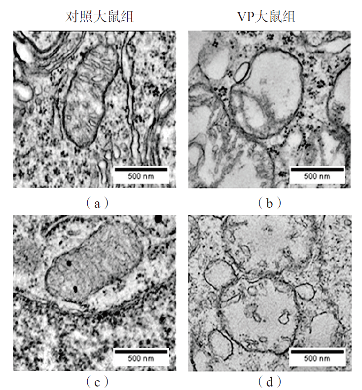
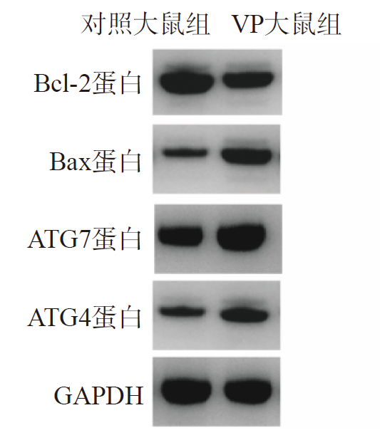
目的 探讨血管性帕金森综合征(VP)患者血清线粒体自噬信号通路PTEN诱导假定激酶1(PINK1)/Parkin相关蛋白的变化,及其对神经功能的作用。方法 选取2020年3月—2022年5月河北北方学院附属第一医院VP患者55例(VP组)、健康志愿者55名(正常对照组)。检测所有研究对象PINK1和Parkin蛋白水平。选取30只SD大鼠,分为对照大鼠组(15只)和VP大鼠组(15只)。采用腹腔注射1-甲基-4-苯基-1,2,3,6-四氢吡啶(MPTP)构建VP大鼠模型,对照大鼠注射等量0.9%NaCl溶液,模型构建成功24 h后采用流式细胞术检测各组大鼠脑组织细胞的存活率、早期凋亡率和晚期凋亡率,采用荧光定量聚合酶链反应(PCR)和免疫印迹法检测大鼠脑组织自噬基因PINK1、Parkin mRNA和蛋白表达情况,同时检测PINK1/Parkin信号通路相关蛋白磷酸化修饰水平[磷酸化PTEN诱导假定激酶1(p-PINK1)蛋白和磷酸化Parkin(p-Parkin)蛋白]。采用透射电镜原位验证大鼠脑组织超微结构的变化情况,并采用免疫印迹法检测自噬蛋白[自噬相关蛋白(ATG)4、ATG7]和凋亡蛋白(Bcl-2、Bax)的相对表达量。结果 VP组血清PINK1和Parkin水平显著高于正常对照组(P<0.05)。与对照大鼠组比较,VP大鼠组脑组织细胞存活率显著降低(P<0.01),细胞早期凋亡率和晚期凋亡率显著升高(P<0.01);PINK1 mRNA和Parkin mRNA相对表达量显著升高(P<0.01),PINK1蛋白、Parkin蛋白、p-PINK1蛋白和p-Parkin蛋白相对表达量均显著升高(P<0.01)。透射电镜分析结果显示,VP大鼠组出现明显的细胞自噬和线粒体自噬损伤。与对照大鼠组比较,VP大鼠组脑组织Bcl-2蛋白相对表达量降低(P<0.001),Bax蛋白、ATG4蛋白和ATG7蛋白相对表达量均升高(P<0.01)。结论 VP会引起脑组织细胞自噬增加,并加重线粒体自噬损伤,与脑组织PINK1/Parkin信号通路的过度激活有关。

关键词: PTEN诱导假定激酶1/Parkin信号通路, 线粒体, 自噬损伤, 血管性帕金森综合征

Abstract:

Objective To investigate the changes in serum levels of PTEN-induced putative kinase 1(PINK1)/Parkin related proteins induced by mitochondrial autophagy signaling pathway in patients with vascular Parkinsonism(VP) and its effect on neural function. Methods Totally,55 VP patients(VP group) and 55 healthy subjects(healthy control group) were enrolled from the First Affiliated Hospital of Hebei North University from March 2020 to May 2022. PINK1 and Parkin protein levels were determined. A total of 30 SD rats were selected and classified into control group(15 rats) and VP group(15 rats). The VP rat model was constructed by intraperitoneal injection of 1-methyl-4-phenyl-1,2,3,6-tetrahydropyridine(MPTP),and the control group was injected with the same amount of 0.9% NaCl solution. For 24 h after the successful construction of the VP model,the survival rate,early apoptosis rate and late apoptosis rate of rat brain tissue were detected by flow cytometry. The mRNA and protein expressions of autophagy genes(PINK1 and Parkin) in rat brain tissue were detected by fluorescence quantitative polymerase chain reaction(PCR) and western blot. Simultaneously,the level of phosphorylation modification of PINK1/Parkin signaling pathway was detected [phosphorylation of PTEN-induced putative kinase 1(p-PINK1) protein and phosphorylation of Parkin(p-Parkin) protein]. The changes of brain ultrastructure were verified in situ by transmission electron microscopy,and the relative expression levels of autophagy proteins [autophagy-related gene(ATG)4 and ATG7] and apoptotic molecules(Bcl-2 and Bax) were detected by western blot. Results The serum PINK1 and Parkin levels in VP group were higher than those in healthy control group(P<0.01). Compared with the control group,the survival rate of VP rats was decreased(P<0.01),and the early and late apoptosis rates were increased(P<0.01). The relative expressions of PINK1 mRNA and Parkin mRNA were increased(P<0.01),and the relative expressions of PINK1 protein,Parkin protein,p-PINK1 protein and p-Parkin protein were increased(P<0.01). The results of transmission electron microscopy showed that autophagy and mitochondrial autophagy were obviously damaged in VP rats. Compared with the control group,the relative expression of Bcl-2 protein in VP rats was decreased(P<0.001),and the relative expressions of Bax,ATG4 and ATG7 proteins were increased(P<0.01). Conclusions VP can increase brain autophagy and aggravate mitochondrial autophagy damage,which is related to the overactivation of brain autophagy signaling pathway PINK1/Parkin.

Key words: PTEN-induced putative kinase 1/Parkin signaling pathway, Mitochondria, Autophagy damage, Vascular Parkinsonism

中图分类号: