
检验医学 ›› 2026, Vol. 41 ›› Issue (1): 20-27.DOI: 10.3969/j.issn.1673-8640.2026.01.004
尹秀杉1, 谭雪玲1, 何仁栋2, 邢艳3( )
)
收稿日期:2024-09-24
修回日期:2025-04-13
出版日期:2026-01-30
发布日期:2026-01-30
通讯作者:
邢艳
作者简介:邢 艳,E-mail:xingy@nsmc.edu.cn。基金资助:
YIN Xiushan1, TAN Xueling1, HE Rendong2, XING Yan3()
Received:2024-09-24
Revised:2025-04-13
Online:2026-01-30
Published:2026-01-30
Contact:
XING Yan
摘要:
目的 探讨系统性红斑狼疮(SLE)患者外周血单核细胞亚群与病情的相关性。方法 选取2022年1—8月川北医学院附属医院SLE患者67例(SLE组)、健康体检者46名(正常对照组)。收集所有研究对象的年龄、性别等一般资料和单核细胞百分比(MO%)、单核细胞亚群[经典型单核细胞百分比(CM%)、中间型单核细胞百分比(IM%)、非经典型单核细胞百分比(NCM%)],以及SLE患者的疾病活动性指标[单核细胞绝对数(MO#)、红细胞沉降率(ESR)、C反应蛋白(CRP)、补体(C)3、C4、抗双链DNA(dsDNA)抗体、抗核抗体(ANA)]检测结果。采用Spearman秩相关分析评估SLE患者MO#、CM%、IM%与疾病活动性指标的相关性。采用Logistic回归分析评估SLE疾病活动性的影响因素。采用受试者工作特征(ROC)曲线评价CM%和IM%诊断SLE的效能。结果 与正常对照组比较,SLE组MO#、MO%、IM%升高(P<0.001),CM%降低(P<0.001),NCM%差异无统计学意义(P>0.05);SLE缓解组MO#、MO%、IM%升高(P<0.05),CM%、NCM%差异无统计学意义(P>0.05)。与SLE缓解组比较,SLE活动组CM%降低(P<0.001),IM%升高(P<0.001),MO#、MO%、NCM%差异均无统计学意义(P>0.05)。SLE活动组CM%与CRP、抗dsDNA抗体水平均呈负相关(P<0.05),与C3、C4水平均呈正相关(P<0.05);IM%与CRP、抗dsDNA抗体水平均呈正相关(P<0.05),与C4水平呈负相关(P<0.05)。年龄较小和IM%升高是SLE患者疾病活动性的危险因素[比值比(OR)值分别为0.948、2.410,95%可信区间(CI)分别为0.900~0.998、1.386~4.192,P<0.05]。CM%和IM%诊断SLE的曲线下面积(AUC)分别为0.785、0.827,判断SLE疾病活动性的AUC分别为0.951、0.966。结论 单核细胞亚群分布与SLE疾病活动性密切相关,或可作为SLE诊断和疾病活动性评估的新的生物标志物。
中图分类号:
尹秀杉, 谭雪玲, 何仁栋, 邢艳. 系统性红斑狼疮患者单核细胞亚群分布与病情的相关性[J]. 检验医学, 2026, 41(1): 20-27.
YIN Xiushan, TAN Xueling, HE Rendong, XING Yan. Correlation between monocyte subset distribution in patients with systemic lupus erythematosus and disease status[J]. Laboratory Medicine, 2026, 41(1): 20-27.
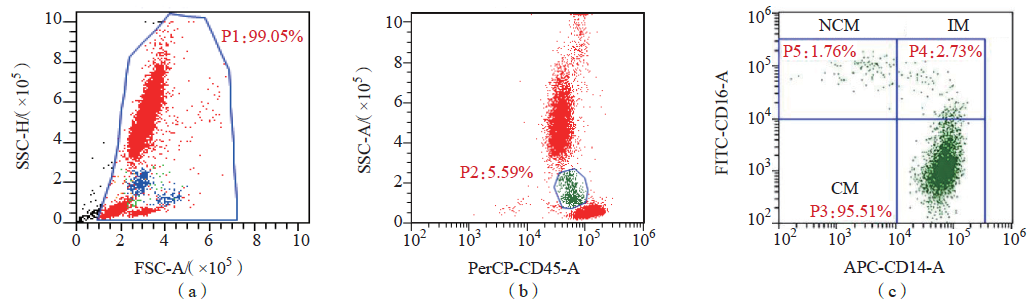
图1 流式细胞术检测MO%和单核细胞亚群的设门策略 注:(a)有核细胞(P1)设门;(b)单核细胞(P2,MO%)设门;(c)不同亚型的单核细胞[P3为CM亚群(CD14++CD16-),P4为IM亚群(CD14++CD16+),P5为NCM亚群(CD14+CD16++)]。
| 组别 | 例数 | 性别 | 年龄/岁 | ESR/(mm·h-1) | CRP/(mg·L-1) | |
|---|---|---|---|---|---|---|
| 男/例 | 女/例 | |||||
| SLE活动组 | 33 | 3 | 30 | 39.50(30.75,51.25) | 14(10,22) | 2.07(0.68,5.63) |
| SLE缓解组 | 34 | 4 | 30 | 41.00(33.25,52.25) | 8(2,18) | 0.76(0.29,2.07) |
| 正常对照组 | 46 | 4 | 42 | 41.07(31.50,52.00) | ||
| 统计值 | 0.232 | 2.592 | 370.50 | 217.50 | ||
| P值 | 0.891 | 0.274 | 0.016 | 0.037 | ||
| 组别 | C3/(mg·L-1) | C4/(mg·L-1) | 抗dsDNA抗体/(IU·mL-1) | ANA滴度 | ||
| SLE活动组 | 753.0(653.5,879.0) | 161.0(123.0,244.0) | 15.84(2.94,63.38) | 1(0,2) | ||
| SLE缓解组 | 756.5(679.8,882.3) | 171.0(127.3,224.5) | 1.70(1.00,11.70) | 1(0,1) | ||
| 正常对照组 | ||||||
| 统计值 | 525.00 | 546.50 | 312.50 | 474.00 | ||
| P值 | 0.656 | 0.856 | 0.002 | 0.246 | ||
表1 SLE活动组、SLE缓解组和正常对照组一般资料和疾病活动性指标检测结果比较
| 组别 | 例数 | 性别 | 年龄/岁 | ESR/(mm·h-1) | CRP/(mg·L-1) | |
|---|---|---|---|---|---|---|
| 男/例 | 女/例 | |||||
| SLE活动组 | 33 | 3 | 30 | 39.50(30.75,51.25) | 14(10,22) | 2.07(0.68,5.63) |
| SLE缓解组 | 34 | 4 | 30 | 41.00(33.25,52.25) | 8(2,18) | 0.76(0.29,2.07) |
| 正常对照组 | 46 | 4 | 42 | 41.07(31.50,52.00) | ||
| 统计值 | 0.232 | 2.592 | 370.50 | 217.50 | ||
| P值 | 0.891 | 0.274 | 0.016 | 0.037 | ||
| 组别 | C3/(mg·L-1) | C4/(mg·L-1) | 抗dsDNA抗体/(IU·mL-1) | ANA滴度 | ||
| SLE活动组 | 753.0(653.5,879.0) | 161.0(123.0,244.0) | 15.84(2.94,63.38) | 1(0,2) | ||
| SLE缓解组 | 756.5(679.8,882.3) | 171.0(127.3,224.5) | 1.70(1.00,11.70) | 1(0,1) | ||
| 正常对照组 | ||||||
| 统计值 | 525.00 | 546.50 | 312.50 | 474.00 | ||
| P值 | 0.656 | 0.856 | 0.002 | 0.246 | ||
| 组别 | 例数 | MO#/(×109L-1) | MO%/% | CM%/% | IM%/% | NCM%/% |
|---|---|---|---|---|---|---|
| 正常对照组 | 46 | 0.29(0.24,0.34) | 5.37(4.85,6.66) | 95.81(94.06,97.05) | 1.97(1.35,3.32) | 1.87(1.11,2.67) |
| SLE组 | 67 | 0.37(0.30,0.52) | 7.97(6.20,9.72) | 92.31(88.38,94.45) | 5.79(3.47,8.33) | 1.93(1.02,2.71) |
| U值 | 758.50 | 663.50 | 662.00 | 532.50 | 1 540.00 | |
| P值 | <0.001 | <0.001 | <0.001 | <0.001 | 0.995 |
表2 SLE组和正常对照组外周血单核细胞及其亚群比较
| 组别 | 例数 | MO#/(×109L-1) | MO%/% | CM%/% | IM%/% | NCM%/% |
|---|---|---|---|---|---|---|
| 正常对照组 | 46 | 0.29(0.24,0.34) | 5.37(4.85,6.66) | 95.81(94.06,97.05) | 1.97(1.35,3.32) | 1.87(1.11,2.67) |
| SLE组 | 67 | 0.37(0.30,0.52) | 7.97(6.20,9.72) | 92.31(88.38,94.45) | 5.79(3.47,8.33) | 1.93(1.02,2.71) |
| U值 | 758.50 | 663.50 | 662.00 | 532.50 | 1 540.00 | |
| P值 | <0.001 | <0.001 | <0.001 | <0.001 | 0.995 |
| 组别 | 例数 | MO#/(×109L-1) | MO%/% | CM%/% | IM%/% | NCM%/% |
|---|---|---|---|---|---|---|
| SLE活动组 | 33 | 0.37(0.30,0.50) | 7.97(5.98,10.93) | 90.66(85.02,92.30) | 7.89(5.82,10.27) | 2.12(1.26,2.60) |
| SLE缓解组 | 34 | 0.39(0.29,0.54) | 8.03(6.30,9.35) | 94.26(92.30,97.05) | 3.64(1.63,5.80) | 1.42(0.93,2.83) |
| U值 | 544.50 | 558.00 | 188.50 | 171.00 | 473.00 | |
| P值 | 0.836 | 0.970 | <0.001 | <0.001 | 0.270 |
表3 SLE缓解组和SLE活动组外周血单核细胞及其亚群比较
| 组别 | 例数 | MO#/(×109L-1) | MO%/% | CM%/% | IM%/% | NCM%/% |
|---|---|---|---|---|---|---|
| SLE活动组 | 33 | 0.37(0.30,0.50) | 7.97(5.98,10.93) | 90.66(85.02,92.30) | 7.89(5.82,10.27) | 2.12(1.26,2.60) |
| SLE缓解组 | 34 | 0.39(0.29,0.54) | 8.03(6.30,9.35) | 94.26(92.30,97.05) | 3.64(1.63,5.80) | 1.42(0.93,2.83) |
| U值 | 544.50 | 558.00 | 188.50 | 171.00 | 473.00 | |
| P值 | 0.836 | 0.970 | <0.001 | <0.001 | 0.270 |
| 项目 | ESR | CRP | C3 | C4 | 抗dsDNA抗体 | ANA |
|---|---|---|---|---|---|---|
| SLE活动组 | ||||||
| CM% | ||||||
| r值 | -0.165 0 | -0.383 1 | 0.388 0 | 0.850 1 | -0.766 2 | -0.082 7 |
| P值 | 0.358 8 | 0.037 2 | 0.025 6 | 0.000 1 | 0.000 1 | 0.647 5 |
| IM% | ||||||
| r值 | 0.173 4 | 0.370 5 | -0.255 2 | -0.834 1 | 0.710 7 | -0.178 2 |
| P值 | 0.334 5 | 0.043 9 | 0.151 7 | 0.000 1 | 0.000 1 | 0.321 0 |
| SLE缓解组 | ||||||
| CM% | ||||||
| r值 | -0.273 7 | -0.162 7 | -0.340 9 | -0.015 3 | -0.146 7 | 0.066 2 |
| P值 | 0.117 3 | 0.469 4 | 0.048 5 | 0.931 6 | 0.407 8 | 0.710 0 |
| IM% | ||||||
| r值 | 0.336 9 | 0.132 8 | 0.277 9 | -0.087 6 | 0.032 9 | -0.063 7 |
| P值 | 0.051 4 | 0.555 9 | 0.111 5 | 0.622 4 | 0.853 6 | 0.720 4 |
表4 SLE患者CM%、IM%与疾病活动性指标的相关性
| 项目 | ESR | CRP | C3 | C4 | 抗dsDNA抗体 | ANA |
|---|---|---|---|---|---|---|
| SLE活动组 | ||||||
| CM% | ||||||
| r值 | -0.165 0 | -0.383 1 | 0.388 0 | 0.850 1 | -0.766 2 | -0.082 7 |
| P值 | 0.358 8 | 0.037 2 | 0.025 6 | 0.000 1 | 0.000 1 | 0.647 5 |
| IM% | ||||||
| r值 | 0.173 4 | 0.370 5 | -0.255 2 | -0.834 1 | 0.710 7 | -0.178 2 |
| P值 | 0.334 5 | 0.043 9 | 0.151 7 | 0.000 1 | 0.000 1 | 0.321 0 |
| SLE缓解组 | ||||||
| CM% | ||||||
| r值 | -0.273 7 | -0.162 7 | -0.340 9 | -0.015 3 | -0.146 7 | 0.066 2 |
| P值 | 0.117 3 | 0.469 4 | 0.048 5 | 0.931 6 | 0.407 8 | 0.710 0 |
| IM% | ||||||
| r值 | 0.336 9 | 0.132 8 | 0.277 9 | -0.087 6 | 0.032 9 | -0.063 7 |
| P值 | 0.051 4 | 0.555 9 | 0.111 5 | 0.622 4 | 0.853 6 | 0.720 4 |
| 项目 | β值 | 标准误 | Wald值 | P值 | OR值(95%CI) |
|---|---|---|---|---|---|
| 性别 | 0.196 | 1.031 | 0.036 | 0.849 | 1.216(0.161~9.170) |
| 年龄 | -0.053 | 0.026 | 4.097 | 0.043 | 0.948(0.900~0.998) |
| CM% | 0.223 | 0.170 | 1.708 | 0.191 | 1.249(0.895~1.745) |
| IM% | 0.880 | 0.282 | 9.703 | 0.002 | 2.410(1.386~4.192) |
表5 SLE疾病活动性的危险因素分析
| 项目 | β值 | 标准误 | Wald值 | P值 | OR值(95%CI) |
|---|---|---|---|---|---|
| 性别 | 0.196 | 1.031 | 0.036 | 0.849 | 1.216(0.161~9.170) |
| 年龄 | -0.053 | 0.026 | 4.097 | 0.043 | 0.948(0.900~0.998) |
| CM% | 0.223 | 0.170 | 1.708 | 0.191 | 1.249(0.895~1.745) |
| IM% | 0.880 | 0.282 | 9.703 | 0.002 | 2.410(1.386~4.192) |
| 项目 | AUC (95%CI) | 最佳临界值/% | 敏感性/ % | 特异性/ % | Youden指数 |
|---|---|---|---|---|---|
| CM% | 0.785(0.701~0.869) | 94.47 | 76.12 | 73.91 | 0.500 3 |
| IM% | 0.827(0.752~0.903) | 3.93 | 70.15 | 89.13 | 0.592 8 |
表6 CM%和IM%诊断SLE的效能
| 项目 | AUC (95%CI) | 最佳临界值/% | 敏感性/ % | 特异性/ % | Youden指数 |
|---|---|---|---|---|---|
| CM% | 0.785(0.701~0.869) | 94.47 | 76.12 | 73.91 | 0.500 3 |
| IM% | 0.827(0.752~0.903) | 3.93 | 70.15 | 89.13 | 0.592 8 |
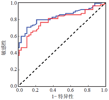
图2 CM%和IM%诊断SLE的ROC曲线 注: CM%; IM%; 参考线。
| 项目 | AUC (95%CI) | 最佳临界值/% | 敏感性/ % | 特异性/ % | Youden指数 |
|---|---|---|---|---|---|
| CM% | 0.951(0.910~0.992) | 92.69 | 90.91 | 89.13 | 0.800 4 |
| IM% | 0.996(0.928~1.000) | 5.05 | 87.88 | 95.65 | 0.835 3 |
表7 CM%和IM%判断SLE活动性的效能
| 项目 | AUC (95%CI) | 最佳临界值/% | 敏感性/ % | 特异性/ % | Youden指数 |
|---|---|---|---|---|---|
| CM% | 0.951(0.910~0.992) | 92.69 | 90.91 | 89.13 | 0.800 4 |
| IM% | 0.996(0.928~1.000) | 5.05 | 87.88 | 95.65 | 0.835 3 |
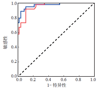
图3 CM%和IM%判断SLE活动性的ROC曲线 注: CM%; IM%; 参考线。
| [1] |
HOI A, IGEL T, MOK C C, et al. Systemic lupus erythematosus[J]. Lancet, 2024, 403(10441):2326-2338.
DOI PMID |
| [2] |
RAMOS-MARTÍNEZ I, RAMOS-MARTÍNEZ E, CERBÓN M, et al. The role of B cell and T cell glycosylation in systemic lupus erythematosus[J]. Int J Mol Sci, 2023, 24(1):863.
DOI URL |
| [3] |
吕迎霞, 杨俊梅, 郑莉娟, 等. SLE患儿外周血TLR-4、HMGB1的表达及临床意义[J]. 检验医学, 2020, 35(11):1143-1146.
DOI |
| [4] |
ARNAUD L, CHASSET F, MARTIN T. Immunopathogenesis of systemic lupus erythematosus:an update[J]. Autoimmun Rev, 2024, 23(10):103648.
DOI URL |
| [5] |
TORRES-RUIZ J, RULL-GABAYET M, MEJÍA-DOMÍNGUEZ N R, et al. Disease activity is associated with changes in the innate immune function in patients with systemic lupus erythematosus[J]. Clin Rheumatol, 2024, 43(1):501-509.
DOI |
| [6] | ZIEGLER-HEITBROCK L, ANCUTA P, CROWE S, et al. Nomenclature of monocytes and dendritic cells in blood[J]. Blood, 2010, 116(16):e74-e80. |
| [7] |
CORMICAN S, GRIFFIN M D. Human monocyte subset distinctions and function:insights from gene expression analysis[J]. Front Immunol, 2020, 11:1070.
DOI URL |
| [8] |
PRAJZLEROVÁ K, KRYŠTŮFKOVÁ O, KOMARC M, et al. The dysregulation of monocyte subpopulations in individuals at risk of developing rheumatoid arthritis[J]. Rheumatology (Oxford), 2021, 60(4):1823-1831.
DOI PMID |
| [9] |
SMILJANOVIC B, RADZIKOWSKA A, KUCA-WARNAWIN E, et al. Monocyte alterations in rheumatoid arthritis are dominated by preterm release from bone marrow and prominent triggering in the joint[J]. Ann Rheum Dis, 2018, 77(2):300-308.
DOI PMID |
| [10] |
SCHNEIDER L, MARCONDES N A, HAX V, et al. Flow cytometry evaluation of CD14/CD16 monocyte subpopulations in systemic sclerosis patients:a cross sectional controlled study[J]. Adv Rheumatol, 2021, 61(1):27.
DOI |
| [11] |
BURBANO C, VASQUEZ G, ROJAS M. Modulatory effects of CD14+CD16++ monocytes on CD14++CD16- monocytes:a possible explanation of monocyte alterations in systemic lupus erythematosus[J]. Arthritis Rheumatol, 2014, 66(12):3371-3381.
DOI URL |
| [12] | FERRETÉ-BONASTRE A G, MARTÍNEZ-GALLO M, MORANTE-PALACIOS O, et al. Disease activity drives divergent epigenetic and transcriptomic reprogramming of monocyte subpopulations in systemic lupus erythematosus[J]. Ann Rheum Dis, 2024, 83(7):865-878. |
| [13] |
ARINGER M, COSTENBADER K, JOHNSON S R. Assessing the EULAR/ACR classification criteria for patients with systemic lupus erythematosus[J]. Expert Rev Clin Immunol, 2022, 18(2):135-144.
DOI URL |
| [14] |
杨玉嘉, 盛家艺, 王潇, 等. 单核细胞分布宽度诊断成人脓毒症准确性系统评价Meta分析[J]. 检验医学, 2024, 39(8):800-806.
DOI |
| [15] |
DASH S P, GUPTA S, SARANGI P P. Monocytes and macrophages:origin,homing,differentiation,and functionality during inflammation[J]. Heliyon, 2024, 10(8):e29686.
DOI URL |
| [16] |
MEDRANO-BOSCH M, SIMÓN-CODINA B, JIMÉNEZ W, et al. Monocyte-endothelial cell interactions in vascular and tissue remodeling[J]. Front Immunol, 2023, 14:1196033.
DOI URL |
| [17] |
GUILLIAMS M, MILDNER A, YONA S. Developmental and functional heterogeneity of monocytes[J]. Immunity, 2018, 49(4):595-613.
DOI PMID |
| [18] | TEH Y C, CHOOI M Y, CHONG S Z. Behind the monocyte's mystique:uncovering their developmental trajectories and fates[J]. Discov Immunol, 2023, 2(1):kyad008. |
| [19] |
RUDER A V, WETZELS S M, TEMMERMAN L, et al. Monocyte heterogeneity in cardiovascular disease[J]. Cardiovasc Res, 2023, 119(11):2033-2045.
DOI PMID |
| [20] | PEREIRA V I C, DE BRITO JUNIOR L C, FALCÃO L F M, et al. Monocytes subpopulations pattern in the acute respiratory syndrome coronavirus 2 virus infection and after long COVID-19[J]. Int Immunopharmacol, 2023, 124(Pt B):110994. |
| [21] |
WU Z, ZHANG S, ZHAO L, et al. Upregulation of CD16- monocyte subsets in systemic lupus erythematous patients[J]. Clin Rheumatol, 2017, 36(10):2281-2287.
DOI PMID |
| [22] |
JIANG W, ZHANG L, LANG R, et al. Sex differences in monocyte activation in systemic lupus erythematosus (SLE)[J]. PLoS One, 2014, 9(12):e114589.
DOI URL |
| [23] | SANTACRUZ J C, MANTILLA M J, RUEDA I, et al. A practical perspective of the hematologic manifestations of systemic lupus erythematosus[J]. Cureus, 2022, 14(3):e22938. |
| [24] |
DUROUX-RICHARD I, ROBIN M, PEILLEX C, et al. MicroRNAs:fine tuners of monocyte heterogeneity[J]. Front Immunol, 2019, 10(9):2145.
DOI URL |
| [25] | TRZEBANSKI S, JUNG S. Plasticity of monocyte development and monocyte fates[J]. Immunol Lett, 2020,227:66-78. |
| [26] |
WILLIAMS H, MACK C, BARAZ R, et al. Monocyte differentiation and heterogeneity:inter-subset and interindividual differences[J]. Int J Mol Sci, 2023, 24(10):8757.
DOI URL |
| [27] |
OŻAŃSKA A, SZYMCZAK D, RYBKA J. Pattern of human monocyte subpopulations in health and disease[J]. Scand J Immunol, 2020, 92(1):e12883.
DOI URL |
| [28] | OHTEKI T. Identification of a human progenitor strictly committed to monocytic differentiation:a counterpart of mouse cMoPs[J]. Rinsho Ketsueki, 2018, 59(6):812-818. |
| [29] |
HAMON P, LOYHER P L, BAUDESSON DE CHANVILLE C, et al. CX3CR1-dependent endothelial margination modulates Ly6Chigh monocyte systemic deployment upon inflammation in mice[J]. Blood, 2017, 129(10):1296-1307.
DOI URL |
| [30] |
MEGHRAOUI-KHEDDAR A, BARTHELEMY S, BOISSONNAS A, et al. Revising CX3CR1 expression on murine classical and non-classical monocytes[J]. Front Immunol, 2020, 11:1117.
DOI URL |
| [31] | 刘讷敏. 系统性红斑狼疮患者外周血中单核细胞亚群与病情活动的相关性研究[D]. 广州: 广州医科大学, 2018. |
| [32] | 袁佳仪, 王岚, 徐学静, 等. 外周血单核细胞亚群分布与类风湿关节炎发病相关性[J]. 中华检验医学杂志, 2022, 45(9):906-913. |
| [33] |
TORRES-RUIZ J, CARRILLO-VAZQUEZ D A, PADILLA-ORTIZ D M, et al. TLR expression in peripheral monocyte subsets of patients with idiopathic inflammatory myopathies:association with clinical and immunological features[J]. J Transl Med, 2020, 18(1):125.
DOI |
| [34] |
ORTEGA MORENO L, FERNÁNDEZ-TOMÉ S, CHAPARRO M, et al. Profiling of human circulating dendritic cells and monocyte subsets discriminates between type and mucosal status in patients with inflammatory bowel disease[J]. Inflamm Bowel Dis, 2021, 27(2):268-274.
DOI PMID |
| [35] |
JHA A, JOSEPH J, PRABHU S B, et al. Utility of peripheral blood monocyte subsets,circulating immune complexes and serum cytokines in assessment of SLE activity:an observational,cross-sectional study[J]. Clin Rheumatol, 2024, 43(1):209-217.
DOI |
| [1] | 李飞, 易长林, 金佩佩, 王芳, 丁宁. 纤维蛋白原/白蛋白比值在SLE疾病活动度和LN诊断中的临床价值[J]. 检验医学, 2025, 40(7): 654-659. |
| [2] | 郑莹, 陆喆, 薛静. SLE患儿血清25(OH)D3水平与淋巴细胞亚群的关系[J]. 检验医学, 2025, 40(3): 230-234. |
| [3] | 刘小敏, 张勇刚, 王素云, 宋锦旗, 张肄鹏. 伴系统性红斑狼疮的MDS-5q- 1例报道[J]. 检验医学, 2021, 36(5): 574-576. |
| [4] | 吕迎霞, 杨俊梅, 郑莉娟, 黄庆华. SLE患儿外周血TLR-4、HMGB1的表达及临床意义[J]. 检验医学, 2020, 35(11): 1143-1146. |
| [5] | 陈艳红, 孙丽, 付昱, 刘艳, 牛国平. 血浆GAS5在系统性红斑狼疮患者诊断及治疗中的临床应用[J]. 检验医学, 2019, 34(8): 696-700. |
| [6] | 王霞, 索明环, 胡婷, 胡耀宗, 温冬梅. IL-33及其受体sST2在SLE患儿血清中的表达及意义[J]. 检验医学, 2019, 34(6): 518-521. |
| [7] | 徐黎明, 沈军. 血清瘦素和脂联素联合检测在狼疮性肾炎诊断中的价值[J]. 检验医学, 2019, 34(5): 401-404. |
| [8] | 李池慧, 郑冰, 俞翀曌. 895例系统性红斑狼疮患者自身抗体检测结果的分析[J]. 检验医学, 2018, 33(5): 463-465. |
| [9] | 谭立明, 焦安君, 冯晓晶, 徐镠粤, 谭福燕, 何思齐, 罗姮, 陈娟娟, 蒋永清, 李华. 抗中性粒细胞胞浆抗体检测对系统性血管炎的临床价值[J]. 检验医学, 2018, 33(2): 101-105. |
| [10] | 俞虹. 系统性红斑狼疮患者血清FER、CA125的检测及临床意义[J]. 检验医学, 2018, 33(2): 182-184. |
| [11] | 宋睿, 叶萍, 陈晓翔. IgG型、IgM型及IgA型抗ds-DNA抗体与SLE的相关性[J]. 检验医学, 2018, 33(11): 969-974. |
| [12] | 陈水绵, 俞翀曌, 李恩灵, 朱昊明, 陆靖波, 郑冰. 自身免疫性疾病患者及健康体检者自身抗体相关实验室指标分析[J]. 检验医学, 2018, 33(1): 31-36. |
| [13] | 谭立明, 丁耀东, 陈娟娟, 李华, 将永清, 万雅妮, 王丽贇, 王田. C1q在自身免疫性疾病中的临床意义[J]. 检验医学, 2017, 32(8): 686-690. |
| [14] | 宋睿, 叶萍, 魏朝晖, 陈晓翔, 王久存. 抗双链DNA抗体与系统性红斑狼疮临床表型的相关性及4种试剂盒效能比较[J]. 检验医学, 2017, 32(4): 316-321. |
| [15] | 徐维家1,李志1,杨婷婷2 ,王波1. 系统性红斑狼疮患者Th17和Th1细胞及其细胞因子水平的变化及临床意义[J]. 检验医学, 2013, 28(5): 396-399. |
| 阅读次数 | ||||||
|
全文 |
|
|||||
|
摘要 |
|
|||||
