
Laboratory Medicine ›› 2020, Vol. 35 ›› Issue (5): 481-486.DOI: 10.3969/j.issn.1673-8640.2020.05.019
Previous Articles Next Articles
JIN Shu1, ZHU Qi2, ZHU Yuan1, YUAN Ya3, CAI Xiaoyao4, ZHANG Ji1, YAN Peiyi1
Received:2019-11-15
Online:2020-05-30
Published:2020-06-17
CLC Number:
JIN Shu, ZHU Qi, ZHU Yuan, YUAN Ya, CAI Xiaoyao, ZHANG Ji, YAN Peiyi. Methylation level and mRNA expression of CpG island of HER2 gene in breast cancer and its adjacent tissues[J]. Laboratory Medicine, 2020, 35(5): 481-486.
Add to citation manager EndNote|Ris|BibTeX
URL: https://www.shjyyx.com/EN/10.3969/j.issn.1673-8640.2020.05.019
| 引物名称 | 序列(5'~3') | 产物大小/bp |
|---|---|---|
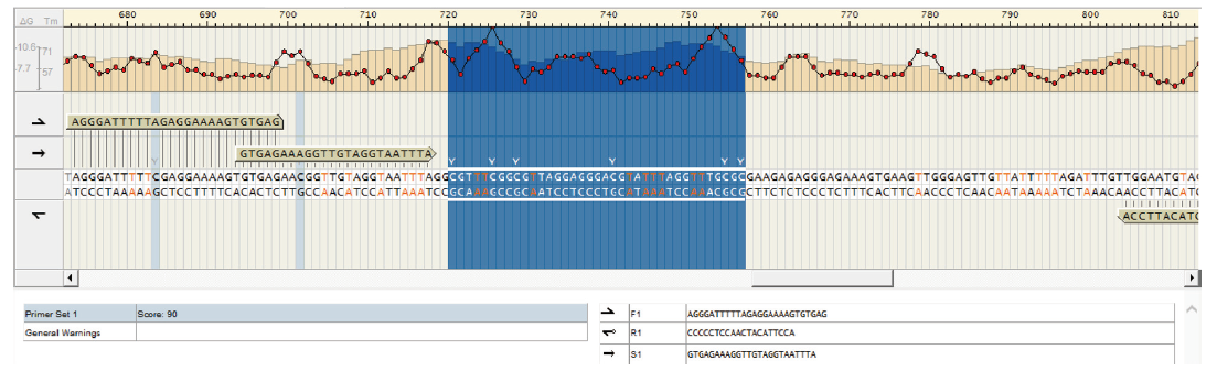
| F1 | AGGGATTTTTAGAGGAAAAGTGTGAG | 151 |
| R1 | CCCCCTCCAACTACATTCCA | |
| S1 | GTGAGAAAGGTTGTAGGTAATTTA |
| 引物名称 | 序列(5'~3') | 产物大小/bp |
|---|---|---|
| F1 | AGGGATTTTTAGAGGAAAAGTGTGAG | 151 |
| R1 | CCCCCTCCAACTACATTCCA | |
| S1 | GTGAGAAAGGTTGTAGGTAATTTA |
| 基因名称 | 序列( 5'~3') | 产物大小/bp |
|---|---|---|
| HER2 | F:GCCCTGTGCCCGAGTGTGCTA R:CAGAAATGCCAGGCTCCCAAAGAT | 124 |
| GAPDH | F:GGATTTGGTCGTATTGGG R:GGAAGATGGTGATGGGATT | 205 |
| 基因名称 | 序列( 5'~3') | 产物大小/bp |
|---|---|---|
| HER2 | F:GCCCTGTGCCCGAGTGTGCTA R:CAGAAATGCCAGGCTCCCAAAGAT | 124 |
| GAPDH | F:GGATTTGGTCGTATTGGG R:GGAAGATGGTGATGGGATT | 205 |
| HER2 | 例数 | HER2 mRNA | 甲基化位点 | 平均甲基化率/% | |||||
|---|---|---|---|---|---|---|---|---|---|
| 1/% | 2/% | 3/% | 4/% | 5/% | 6/% | ||||
| 阳性 | 17 | 0.15(0.05~0.41) | 15.87±6.52 | 14.43±4.46 | 11.66±4.38 | 17.10±5.63 | 17.16±3.67 | 10.60±3.79 | 14.47±4.56 |
| 阴性 | 33 | 0.06(0.02~0.11) | 19.05±10.11 | 15.53±7.09 | 13.33±6.57 | 18.74±8.42 | 17.49±6.05 | 11.46±5.46 | 15.93±7.03 |
| 统计值 | 2.12 | 1.34 | 0.67 | 1.07 | 0.82 | 0.20 | 0.59 | 0.89 | |
| P值 | 0.03 | 0.25 | 0.56 | 0.35 | 0.47 | 0.84 | 0.56 | 0.44 | |
| 项目 | 例数 | HER2 mRNA | 甲基化位点 | 平均甲基化率/% | |||||
|---|---|---|---|---|---|---|---|---|---|
| 1/% | 2/% | 3/% | 4/% | 5/% | 6/% | ||||
| 癌组织 | 17 | 0.15(0.05~0.41) | 15.87±6.52 | 14.43±4.46 | 11.66±4.38 | 17.10±5.63 | 17.16±3.67 | 10.59±3.79 | 14.47±4.57 |
| 癌旁组织 | 17 | 0.15(0.05~0.30) | 13.64±7.98 | 12.06±5.69 | 9.77±5.08 | 14.04±6.59 | 14.92±3.46 | 6.84±3.85 | 11.88±4.67 |
| 统计值 | 0.12 | 0.90 | 1.36 | 1.17 | 1.46 | 1.83 | 2.87 | 1.64 | |
| P值 | 0.90 | 0.38 | 0.19 | 0.25 | 0.16 | 0.08 | 0.01 | 0.11 | |
| 项目 | 例数 | HER2 mRNA | 甲基化位点 | 平均甲基化率/% | |||||
|---|---|---|---|---|---|---|---|---|---|
| 1/% | 2/% | 3/% | 4/% | 5/% | 6/% | ||||
| 癌组织 | 33 | 0.06(0.02~0.11) | 19.05±10.11 | 15.53±7.09 | 13.33±6.57 | 18.74±8.42 | 17.49±6.05 | 11.46±5.46 | 15.93±7.03 |
| 癌旁组织 | 33 | 0.12(0.08~0.18) | 14.85±8.31 | 12.83±5.84 | 10.58±5.65 | 15.04±7.14 | 14.44±6.54 | 7.06±6.24 | 12.75±5.66 |
| 统计值 | 3.53 | 2.16 | 2.02 | 2.11 | 2.19 | 2.62 | 2.86 | 2.23 | |
| P值 | 0.00 | 0.04 | 0.05 | 0.04 | 0.04 | 0.01 | 0.01 | 0.03 | |
| HER2 | 例数 | HER2 mRNA | 甲基化位点 | 平均甲基化率/% | |||||
|---|---|---|---|---|---|---|---|---|---|
| 1/% | 2/% | 3/% | 4/% | 5/% | 6/% | ||||
| 阳性 | 17 | 0.15(0.05~0.30) | 13.64±7.98 | 12.06±5.69 | 9.77±5.08 | 14.04±6.59 | 14.92±3.46 | 6.84±3.85 | 11.88±4.67 |
| 阴性 | 33 | 0.12(0.08~0.18) | 14.85±8.31 | 12.83±5.84 | 10.58±5.65 | 15.04±7.14 | 14.44±6.54 | 7.06±6.24 | 12.75±5.66 |
| 统计值 | 0.090 | 0.49 | 0.45 | 0.50 | 0.48 | -0.28 | 0.15 | 0.54 | |
| P值 | 0.927 | 0.62 | 0.66 | 0.62 | 0.63 | 0.78 | 0.88 | 0.59 | |
| [1] | SIEGEL R L,MILLER K D,JEMAL A.Cancer statistics,2018[J]. CA Cancer J Clin,2018,68(1):7-30. |
| [2] | YAN M,PARKER B A,SCHWAB R,et al.HER2 aberrations in cancer:implications for therapy[J]. Cancer Treat Rev,2014,40(6):770-780. |
| [3] | EKMAN S.HER2:defining a Neu target in non-small-cell lung cancer[J]. Ann Oncol,2019,30(3):353-355. |
| [4] | 金姝,朱琪,秦华晖,等. 乳腺癌C-erbB-2蛋白表达与基因CpG岛甲基化状态和mRNA表达的关系[J]. 检验医学,2011,26(11):750-755. |
| [5] | PEROU C M,SØRLIE T,EISEN M B,et al. Molecular portraits of human breast tumours[J]. Nature,2000,406(6797):747-752. |
| [6] | Cancer Genome Atlas Network. Comprehensive molecular portraits of human breast tumours[J]. Nature,2012,490(7418):61-70. |
| [7] | PARKER J S,MULLINS M,CHEANG M C,et al.Supervised risk predictor of breast cancer based on intrinsic subtypes[J]. J Clin Oncol,2009,27(8):1160-1167. |
| [8] | DABBS D J,KLEIN M E,MOHSIN S K,et al.High false-negative rate of HER2 quantitative reverse transcription polymerase chain reaction of the Oncotype DX test:an independent quality assurance study[J]. J Clin Oncol,2011,29(32):4279-4285. |
| [9] | MOUTTET D,LAÉ M,CALY M,et al.Estrogen-receptor,progesterone-receptor and HER2 status determination in invasive breast cancer. Concordance between immuno-histochemistry and MapQuantTM microarray based assay[J]. PLoS One,2016,11(2):e0146474. |
| [10] | DENKERT C,LOIBL S,KRONENWETT R,et al.RNA-based determination of ESR1 and HER2 expression and response to neoadjuvant chemotherapy[J]. Ann Oncol,2013,24(3):632-639. |
| [11] | KHALED N,BIDET Y.New insights into the implication of epigenetic alterations in the EMT of triple negative breast cancer[J]. Cancers(Basel),2019,11(4):559. |
| [12] | DWORKIN A M,HUANG T H,TOLAND A E.Epigenetic alterations in the breast:implications for breast cancer detection,prognosis and treatment[J]. Semin Cancer Biol,2009,19(3):165-171. |
| [13] | GUY C T,WEBSTER M A,SCHALLER M,et al.Expression of the neu protooncogene in the mammary epithelium of transgenic mice induces metastatic disease[J]. Proc Natl Acad Sci U S A,1992,89(22):10578-10582. |
| [14] | ZHOU H,CHEN W D,QIN X,et al.MMTV promoter hypomethylation is linked to spontaneous and MNU associated c-neu expression and mammary carcinogenesis in MMTV c-neu transgenic mice[J]. Oncogene,2001,20(42):6009-6017. |
| [15] | FREUDENBERG J A,WANG Q,KATSUMATA M,et al.The role of HER2 in early breast cancer metastasis and the origins of resistance to HER2-targeted therapies[J]. Exp Mol Pathol,2009,87(1):1-11. |
| [16] | YAN P S,VENKATARAMU C,IBRAHIM A,et al.Mapping geographic zones of cancer risk with epigenetic biomarkers in normal breast tissue[J]. Clin Cancer Res,2006,12(22):6626-6636. |
| [17] | POLYAK K.Breast cancer:origins and evolution[J]. J Clin Invest,2007,117(11):3155-3163. |
| [18] | DWORKIN A M,HUANG T H,TOLAND A E.Epigenetic alterations in the breast:implications for breast cancer detection,prognosis and treatment[J]. Semin Cancer Biol,2009,19(3):165-171. |
| [19] | SIMONOVA O,KUZNETSOVA E,TANAS A S,et al.Matrix metalloproteinases and their tissue inhibitors genes abnormal DNA methylation in breast cancer[J]. Ann Oncol,2019,30(Suppl 5):v6. |
| [20] | LIU Q,KULAK M V,BORCHERDING N,et al.A novel HER2 gene body enhancer contributes to HER2 expression[J]. Oncogene,2018,37(5):687-694. |
| [21] | LINDQVIST B M,WINGREN S,MOTLAGH P B,et al.Whole genome DNA methylation signature of HER2-positive breast cancer[J]. Epigenetics-US,2014,9(8):1149-1162. |
| [1] | HUANG Ren, SHI Weizhong, LU Renquan, GUO Lin, WANG Yanchun. Combined determination of Septin9 gene methylation and SDC2 gene methylation in auxiliary diagnosis of colorectal cancer [J]. Laboratory Medicine, 2025, 40(6): 519-524. |
| [2] | HUANG Lei, TANG Wenjia, ZHOU Yan, ZHOU Jiaye, ZHANG Chunyan, YANG Jing, WANG Beili, PAN Baishen, GUO wei. Prognosis of RDW-SD and RDW-CV in breast cancer patients with lymphatic metastasis [J]. Laboratory Medicine, 2024, 39(4): 376-381. |
| [3] | LIU Bohan, LIU Yiwen, HE Yiqing, LU Renquan, DU Yan, ZHANG Guoliang, GUO Qian, GAO Feng, YANG Cuixia. Serum amphiregulin and mesothelin in auxiliary diagnosis of breast cancer [J]. Laboratory Medicine, 2024, 39(1): 26-30. |
| [4] | WANG Rong, XING Lianxiang, HUANG Keliang, LI Xin. MiR-374 promoting proliferation and invasion of breast cancer cells by targeting and down-regulating TRIM35 [J]. Laboratory Medicine, 2023, 38(9): 812-817. |
| [5] | CHEN Chen, DUAN Qi, LU Jiatuan, ZHAI Xiaojian, WANG Zheng, ZHANG Hao, GUO Man. Expressions of ASH2L and HOXA2 in triple-negative breast cancer patients and their relationship with lymph node metastasis [J]. Laboratory Medicine, 2023, 38(6): 574-578. |
| [6] | ZHANG Guoliang, LIU Yiwen, HE Yiqing, XU Jing, YANG Cuixia, GAO Feng, LIU Hua. Changes of serum HAS2 and CD44 levels in breast cancer patients and their clinical significance [J]. Laboratory Medicine, 2023, 38(5): 424-429. |
| [7] | GAO Feng. Clinical application of novel tumor biomarkers:prospects and challenges [J]. Laboratory Medicine, 2023, 38(4): 303-306. |
| [8] | LI Mu, GONG Dongliang, XU Liming, PENG Rong. Relationship of XPC rs2228000 polymorphisms and breast cancer [J]. Laboratory Medicine, 2023, 38(3): 235-239. |
| [9] | LIU Chong, ZHANG Jing, LI Sheng, ZHAO Qi. Expressions and correlation of miR-335 and Fra-1 in breast cancer [J]. Laboratory Medicine, 2023, 38(2): 143-147. |
| [10] | GAO Jianchao, WANG Sisi, ZHANG Zhisheng, ZHANG Jingli, LI Xiaoxia, MA Ke, FENG Zhilin, ZHOU Haifeng, WANG Zhanhai. Efficacy and prognosis evaluation of neo-adjuvant chemotherapy for breast cancer based on miR-206,miR-125 and miR-21 [J]. Laboratory Medicine, 2023, 38(11): 1062-1068. |
| [11] | WANG Xiaoye, DONG Guoyou, LIU Zhiying. Relations of ZEB2 and E-Cad expressions in breast cancer tissues with prognosis [J]. Laboratory Medicine, 2022, 37(9): 815-820. |
| [12] | YU Fangfang, ZHAO Qi, YANG Liping, WANG Chenyu. Correlation of hematological indexes and expression of HER-2 in patients with breast cancer [J]. Laboratory Medicine, 2022, 37(6): 514-517. |
| [13] | GONG Zhiyun, JIANG Minglei, SHI Weizhong, LU Renquan, GUO Lin. Application value of fecal SDC2 gene methylation determination in the auxiliary diagnosis of colorectal cancer [J]. Laboratory Medicine, 2022, 37(4): 325-329. |
| [14] | LIU Kai, ZHANG Peiru, XIE Suhong, GUO Lin, LU Renquan. Clinical value of the SorCS1 gene promoter methylation determination in patients with colorectal cancer [J]. Laboratory Medicine, 2022, 37(4): 330-335. |
| [15] | YE Jingwen, SHEN Yunyue, LIU Yiwen, HE Yiqing, DU Yan, ZHANG Guoliang, GAO Feng, YANG Cuixia. Role of MAPK/ERK signaling pathway in reversing endocrine resistance of breast cancer [J]. Laboratory Medicine, 2022, 37(4): 342-348. |
| Viewed | ||||||
|
Full text |
|
|||||
|
Abstract |
|
|||||