
Laboratory Medicine ›› 2015, Vol. 30 ›› Issue (2): 185-190.DOI: 10.3969/j.issn.1673-8640.2015.02.020
• Orignal Article • Previous Articles Next Articles
LI Feng, WEI Qun, HUANG Dongfeng, ZHANG Hon
Received:2014-04-22
Online:2015-02-28
Published:2015-02-12
CLC Number:
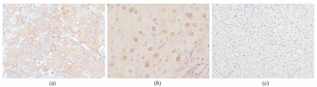
LI Feng, WEI Qun, HUANG Dongfeng, ZHANG Hon. Expressions and significance of gp96 and Mcl-1 in liver cirrhosis and hepatocellular carcinoma tissues[J]. Laboratory Medicine, 2015, 30(2): 185-190.
Add to citation manager EndNote|Ris|BibTeX
URL: https://www.shjyyx.com/EN/10.3969/j.issn.1673-8640.2015.02.020
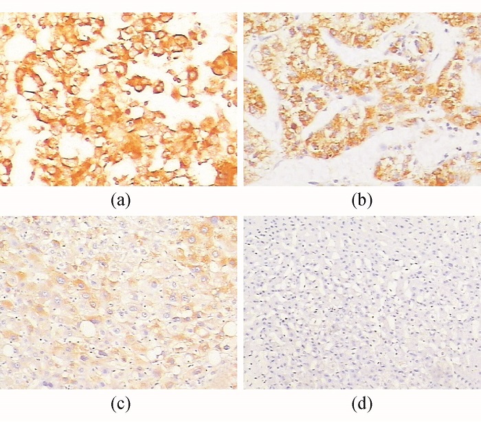
| 组别 | 例数 | gp96 | Mcl-1 | ||
|---|---|---|---|---|---|
| 肝癌组 | 32 | 27 | (84.38)*# | 14 | (43.75)* |
| 肝硬化组 | 19 | 10 | (52.63)* | 6 | (31.58)* |
| 对照组 | 21 | 2 | (9.52) | 1 | (4.76) |
| 组别 | 例数 | gp96 | Mcl-1 | ||
|---|---|---|---|---|---|
| 肝癌组 | 32 | 27 | (84.38)*# | 14 | (43.75)* |
| 肝硬化组 | 19 | 10 | (52.63)* | 6 | (31.58)* |
| 对照组 | 21 | 2 | (9.52) | 1 | (4.76) |
| 组别 | 例数 | gp96 | Mcl-1 |
|---|---|---|---|
| 肝癌组 | 19 | 16(84.21) | 9(47.37) |
| 配对癌旁肝硬化组 | 19 | 10(52.63)* | 6(31.58) |
| 组别 | 例数 | gp96 | Mcl-1 |
|---|---|---|---|
| 肝癌组 | 19 | 16(84.21) | 9(47.37) |
| 配对癌旁肝硬化组 | 19 | 10(52.63)* | 6(31.58) |
| 组别 | 例数 | gp96 | Mcl-1 | |
|---|---|---|---|---|
| 肝癌组 | 13 | 11(84.62) | 6 | (46.15) |
| 配对癌旁非肝硬化组 | 13 | 2(15.38)* | 1 | (7.69)* |
| 组别 | 例数 | gp96 | Mcl-1 | |
|---|---|---|---|---|
| 肝癌组 | 13 | 11(84.62) | 6 | (46.15) |
| 配对癌旁非肝硬化组 | 13 | 2(15.38)* | 1 | (7.69)* |
| 临床病理因素 | 例数 | gp96 | P值 | Mcl-1 | P值 | ||
|---|---|---|---|---|---|---|---|
| + | - | + | - | ||||
| 性别 | 0.198 | 0.740 | |||||
| 男 | 25 | 20 | 5 | 16 | 9 | ||
| 女 | 7 | 7 | 0 | 4 | 3 | ||
| 年龄(岁) | 0.737 | 0.647 | |||||
| <50 | 15 | 13 | 2 | 10 | 5 | ||
| ≥50 | 17 | 14 | 3 | 10 | 7 | ||
| 临床病理因素 | 例数 | gp96 | P值 | Mcl-1 | P值 | ||
|---|---|---|---|---|---|---|---|
| + | - | + | - | ||||
| 性别 | 0.198 | 0.740 | |||||
| 男 | 25 | 20 | 5 | 16 | 9 | ||
| 女 | 7 | 7 | 0 | 4 | 3 | ||
| 年龄(岁) | 0.737 | 0.647 | |||||
| <50 | 15 | 13 | 2 | 10 | 5 | ||
| ≥50 | 17 | 14 | 3 | 10 | 7 | ||
| 临床病理因素 | 例数 | gp96 | P值 | Mcl-1 | P值 | ||
|---|---|---|---|---|---|---|---|
| + | - | + | - | ||||
| 肿瘤大小(cm) | 0.779 | 0.092 | |||||
| ≤3 | 8 | 7 | 1 | 3 | 5 | ||
| >3 | 24 | 20 | 4 | 17 | 7 | ||
| 肿瘤包膜 | 0.002 | 0.102 | |||||
| 有 | 6 | 4 | 2 | 2 | 4 | ||
| 无 | 26 | 26 | 0 | 18 | 8 | ||
| 肿瘤坏死 | 0.160 | 0.040 | |||||
| 有 | 8 | 8 | 0 | 7 | 1 | ||
| 无 | 24 | 19 | 5 | 11 | 13 | ||
| 门静脉癌栓 | 0.198 | 0.151 | |||||
| 有 | 7 | 7 | 0 | 6 | 1 | ||
| 无 | 25 | 20 | 5 | 14 | 11 | ||
| AFP(μg /L) | 0.307 | 0.403 | |||||
| <400 | 13 | 12 | 1 | 7 | 6 | ||
| ≥400 | 19 | 15 | 4 | 13 | 6 | ||
| 组织学分级 | 0.078 | 0.102 | |||||
| Ⅰ~Ⅱ | 21 | 16 | 5 | 11 | 10 | ||
| Ⅲ | 11 | 11 | 0 | 9 | 2 | ||
| 临床分期 | 0.975 | 0.926 | |||||
| Ⅰa~Ⅰb | 13 | 11 | 2 | 8 | 5 | ||
| Ⅱa~Ⅱb | 19 | 16 | 3 | 12 | 7 | ||
| TNM分期 | 0.044 | 0.030 | |||||
| Ⅰ~Ⅱ | 22 | 15 | 7 | 11 | 11 | ||
| Ⅲ | 10 | 10 | 0 | 9 | 1 | ||
| HBsAg | 0.732 | 0.740 | |||||
| + | 25 | 20 | 5 | 16 | 9 | ||
| - | 7 | 6 | 1 | 4 | 3 | ||
| 肝硬化 | 0.975 | 0.946 | |||||
| 有 | 19 | 16 | 3 | 9 | 10 | ||
| 无 | 13 | 11 | 2 | 6 | 7 | ||
| [1] | PARKIN DM,BRAY F,FERLAY J,et al.Global cancer statistics, 2002[J]. CA Cancer J Clin,2005,55(2):74-108. |
| [2] | YANG L,PARKIN DM,FERLAY J,et al.Estimates of cancer incidence in China for 2000 and projections for 2005[J]. Cancer Epidemiol Biolmarkers Prev,2005,14(1):243-250. |
| [3] | LUO RH,ZHAO ZX,ZHOU XY,et al.Risk factors for primary liver carcinoma in Chinese population[J]. World J Gastroenterol,2005,11(28):4431-4434. |
| [4] | HUA Y, WHITE-GILBERTSON S, KELLNER J, et al.Molecular chaperone gp96 is a novel therapeutic target of multiple myeloma[J]. Clin Cancer Res,2013,19(22):6242-6251. |
| [5] | SANO M, NAKANISHI Y, YAGASAKI H, et al.Overexpression of anti-apoptotic Mcl-1 in testicular germ cell tumours[J]. Histopathology,2005,46(5):532-539. |
| [6] | 邢传平,刘斌,董亮. 免疫组织化学标记结果的判断方法[J].中华病理学杂志,2001,30(4):318. |
| [7] | FU Y,LEE AS.Glucose regulated proteins in cancer progression,drug resistance and immunotherapy[J]. Cancer Biol Ther,2006,5(7):741-744. |
| [8] | WANG Q,HE Z,ZHANG J,et al.Overexpression of endoplasmic reticulum molecular chaperone GRP94 and GRP78 in human lung cancer tissues and its significance[J]. Cancer Detect Prev,2005,29(6):544-551. |
| [9] | SINGH-JASUJA H,SCHERER HU,HILF N,et al.The heat shock protein gp96 induces maturation of dendritic cells and down-regulation of its receptor[J]. Eur J Immunol,2000, 30(8):2211-2215. |
| [10] | BELLI F,TESTORI A,RIVOLTINI L,et al.Vaccination of metastatic melanoma patients with autologous tumor-derived heat shock protein gp96-peptide complexes: clinical and immunologic findings[J]. J Clin Oncol,2002,20(20):4169-4180. |
| [11] | MAZZAFERRO V,COPPA J,CARRABBA MG,et al.Vaccination with autologous tumor-derived heat-shock protein gp96 after liver resection for metastatic colorectal cancer[J]. Clin Cancer Res,2003,9(9):3235-3245. |
| [12] | XU Z,JENSEN G,YEN TS.Activation of hepatitis B virus S promoter by the viral large surface protein via induction of stress in the endoplasmic reticulum[J]. J Virol,1997,71(10):7387-7392. |
| [13] | KIM YS,LIM HK,RHIM H,et al.Recurrence of hepatocellular carcinoma after liver transplantation: patterns and prognostic factors based on clinical and radiologic features[J]. AJR AM J Roentgenol,2007,189(2):352-358. |
| [14] | LIU H,PENG HW,CHENG YS,et al.Stabilization and enhancement of the antiapoptotic activity of mcl-1 by TCTP[J]. Mol Cell Biol,2005, 25(8):3117-3126. |
| [15] | SONG L,COPPOLA D,LIVINGSTON S,et al.Mcl-1 regulates survival and sensitivity to diverse apoptotic stimuli in human non-small cell lung cancer cells[J]. Cancer Biol Ther,2005,4(3):267-276. |
| [16] | CHUNG TK,CHEUNG TH,LO WK,et al.Expression of apoptotic regulators and their significance in cervical cancer[J]. Cancer Lett,2002,180(1):63-68. |
| [17] | FLEISCHER B,SCHULZE-BERGKAMEN H,SCHUCHMANN M,et al.Mcl-1 is an anti-apoptotic factor for human hepatocellular carcinoma[J]. Int J Oncol,2006,28(1):25-32. |
| [18] | SIEGHART W,LOSERT D,STROMMER S,et al.Mcl-1 overexpression in hepatocellular carcinoma: a potential target for antisense therapy[J]. J Hepatol,2006,44(1):151-157. |
| [19] | CRAIG RW.MCL1 provides a window on the role of the BCL2 family in cell proliferation,differentiation and tumorigenesis[J]. Leukemia,2002,16(4):444-454. |
| [20] | EDWARDS SW, DEROUET M, HOWSE M, et al.Regulation of neutrophil apoptosis by Mcl-1[J]. Biochem Soc Trans,2004,32(Pt3):489-492. |
| [1] | MA Xiaolu, GUO Lin, LU Renquan. Clinical value and application prospect of circulating biomarkers in hepatocellular carcinoma patients [J]. Laboratory Medicine, 2025, 40(4): 309-316. |
| [2] | TIAN Ze, LIU Hongrui, SI Wenzhe. Research progress of exosomal miRNA as biomarkers of hepatocellular carcinoma [J]. Laboratory Medicine, 2025, 40(4): 317-323. |
| [3] | SUN Haiqing, LIU Ning, LOU Jinli, YU Yanhua. Clinical role of STIP1 and AFP-L3 combined determination in diagnosing HCC [J]. Laboratory Medicine, 2025, 40(4): 324-330. |
| [4] | ZHU Jing, ZHANG Rulin, WU Jun. Expressions of CCNB1,PTTG1 and CBX3 in hepatocellular carcinoma and their roles in prognostic assessment [J]. Laboratory Medicine, 2025, 40(4): 331-337. |
| [5] | WANG Xiaolong, CHEN Yuanbin. Relationship between serum cell division cyclin 42 and clinicopathological characteristics and prognosis of AFP-negative hepatocellular carcinoma patients [J]. Laboratory Medicine, 2025, 40(3): 259-263. |
| [6] | LIU Shanfeng, GAO Yun, WANG Limin, WANG Ping. Accuracy analysis of urine protein and urine cast of hospitalized liver cirrhosis patients [J]. Laboratory Medicine, 2025, 40(3): 287-293. |
| [7] | WANG Tian, ZHANG Yun, BAI Yi, ZHAI Weibin, ZHAO Hai. Roles of PIV,SII and NPAR in the diagnosis of spontaneous bacterial peritonitis in patients with liver cirrhosis and ascites [J]. Laboratory Medicine, 2025, 40(12): 1153-1158. |
| [8] | LIU Yang, HE Chengshan, JIANG Xiudi, LU Zhicheng. HBV PreS/S region gene mutation inducing hepatocyte endoplasmic reticulum stress causing hepatocellular carcinoma [J]. Laboratory Medicine, 2024, 39(12): 1229-1233. |
| [9] | ZHANG Yang, ZHANG Dehe, TANG Shiyue, ZHANG Jun, WANG Jianming, CHEN Ling. Predictive value of soluble CD163 combined with Charlson index for esophageal variceal bleeding in patients with liver cirrhosis [J]. Laboratory Medicine, 2024, 39(1): 19-25. |
| [10] | Clinical Laboratory Society of Chinese Association for Rehabilitation Medicine , Molecular Diagnostics Society of Shanghai Medical Association , Tumor Immunology Branch of Shanghai Society for Immunology , Yueyang Hospital of Integrated Traditional Chinese and Western Medicine of Shanghai University of Traditional Chinese Medicine , Shanghai Center for Clinical Laboratory, Clinical Laboratory Society of Chinese Association of Integrative Medicine , Clinical Laboratory Society of Shanghai Anticancer Association , Tumor Markers Society of Shanghai Anticancer Association . Expert consensus on the clinical application of AFP,AFP-L3% and DCP using GALAD and GALAD-like models in hepatocellular carcinoma [J]. Laboratory Medicine, 2023, 38(7): 607-623. |
| [11] | CHEN Wenju, ZHOU Yong, XU Jiajia, WANG Pan. Role of serum exosomal miR-23b-3p and miR-4429 in patients with hepatocellular carcinoma [J]. Laboratory Medicine, 2023, 38(7): 624-628. |
| [12] | GU Yi, WANG Rui, DENG Jie, YANG Yijing, ZHOU Wangke, CHEN Yunuo, CHEN Xiaosong, SHEN Wei, ZHOU Jingyi. Differential analysis and prognostic diagnostic value of inflammatory biomarkers in liver transplantation in patients with severe hepatitis and cirrhosis [J]. Laboratory Medicine, 2023, 38(11): 1044-1051. |
| [13] | WU Sujun, JI Hengtao, PENG Mengle. Clinical values of IL-18,IL-37,NLRP3 and NK/DC ratio in hepatitis B-related liver cirrhosis [J]. Laboratory Medicine, 2022, 37(6): 518-523. |
| [14] | MENG Jun, WANG Junqing, FEI Xiaochun, GU Zhidong. Establishment and validation of a plasma exosome-derived circular RNA model for HCC diagnosis [J]. Laboratory Medicine, 2022, 37(1): 1-10. |
| [15] | LIU Tingting, LIN Yuting, Mila , LI Xiaoqin. Correlation between serum IL-33 level and its gene polymorphism and the clinical outcome of HBV infection [J]. Laboratory Medicine, 2021, 36(11): 1101-1105. |
| Viewed | ||||||
|
Full text |
|
|||||
|
Abstract |
|
|||||