| 1 |
REYA T,MORRISON SJ,CLAREE MF,et al.Stem cells,cancer,and cancer stem cells[J]. Nature,2001,414(6859):105-111.
|
| 2 |
CHUTHAPISITH S,EREMIN J,EL-SHEEMEY M,et al.Breast cancer chemoresistance:emerging importance of cancer stem cells[J]. Surg Oncol,2010,19(1):27-32.
|
| 3 |
SIEGEL R,NAISHADHAM D,JEMAL A.Cancer statistics,2013[J]. CA Cancer J Clin,2013,63(1):11-30.
|
| 4 |
ZHAO X,HE W,LI J,et al.MiRNA-125b inhibits proliferation and migration by targeting SphK1 in bladder cancer[J]. Am J Transl Res,2015,7(11):2346-2354.
|
| 5 |
LEE M,KIM EJ,JEON MJ.MicroRNAs 125a and 125b inhibit ovarian cancer cells through post-transcriptional inactivation of EIF4EBP1[J]. Oncotarget,2016,7(8):8726-8742.
|
| 6 |
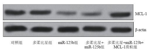
HE H,TIAN W,CHEN H,et al.MicroRNA-101 sensitizes hepatocellular carcinoma cells to doxorubicin-induced apoptosis via targeting Mcl-1[J]. Mol Med Rep,2016,13(2):1923-1929.
|
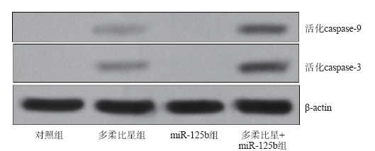
| 7 |
SHUHENDLER AJ,PRASAD P,ZHANG RX,et al.Synergistic nanoparticulate drug combination overcomes multidrug resistance,increases efficacy,and reduces cardiotoxicity in a nonimmunocompromised breast tumor model[J]. Mol Pharm,2014,11(8):2659-2674.
|
| 8 |
KWON T,BAK Y,PARK YH,et al.Peroxiredoxin Ⅱ is essential for maintaining stemness by redox regulation in liver cancer cells[J]. Stem Cells,2016,34(5):1188-1197.
|
| 9 |
SUN JG,XIANG J,ZENG XL,et al.Clitocine induces apoptosis and enhances the lethality of ABT-737 in human colon cancer cells by disrupting the interaction of Mcl-1 and Bak[J]. Cancer Lett,2014,355(2):253-263.
|
| 10 |
PRATHAPAN A,VINEETHA VP,RAGHU KG.Protective effect of Boerhaavia diffusa L. against mitochondrial dysfunction in angiotensin Ⅱ induced hypertrophy in H9c2 cardiomyoblast cells[J]. PLoS One,2014,9(4):e96220.
|
| 11 |
ZHANG HL,WANG P,LU MZ,et al.c-Myc regulation of ATP-binding cassette transporter reverses chemoresistance in CD133(+) colon cancer stem cells[J]. Sheng Li Xue Bao,2016,68(2):171-178.
|
| 12 |
MOHAMMED MK,SHAO C,WANG J,et al.Wnt/β-catenin signaling plays an ever-expanding role in stem cell self-renewal,tumorigenesis and cancer chemoresistance[J]. Genes Dis,2016,3(1):11-40.
|
| 13 |
RAHMANI M,AUST MM,BENSON EC,et al.PI3K/mTOR inhibition markedly potentiates HDAC inhibitor activity in NHL cells through BIM- and MCL-1-dependent mechanisms in vitro and in vivo[J]. Clin Cancer Res,2014,20(18):4849-4860.
|
| 14 |
BEEKMAN AM,HOWELL LA.Small-molecule and peptide inhibitors of the pro-survival protein Mcl-1[J]. ChemMedChem,2016,11(8):802-813.
|
| 15 |
PEI XY,DAI Y,FELTHOUSEN J,et al.Circumvention of Mcl-1-dependent drug resistance by simultaneous Chk1 and MEK1/2 inhibition in human multiple myeloma cells[J]. PLoS One,2014,9(3):e89064.
|
| 16 |
WILLIAMS MM,COOK RS.Bcl-2 family proteins in breast development and cancer:could Mcl-1 targeting overcome therapeutic resistance?[J]. Oncotarget,2015,6(6):3519-3530.
|
| 17 |
JIA HY,WANG YX,YAN WT,et al.MicroRNA-125b functions as a tumor suppressor in hepatocellular carcinoma cells[J]. Int J Mol Sci,2012,13(7):8762-8774.
|
| 18 |
XIE X,HU Y,XU L,et al.The role of miR-125b-mitochondria-caspase-3 pathway in doxorubicin resistance and therapy in human breast cancer[J]. Tumour Biol,2015,36(9):7185-7194.
|
| 19 |
ZHAO L,WANG W. miR-125b suppresses the proliferation of hepatocellular carcinoma cells by targeting Sirtuin7[J]. Int J Clin Exp Med,2015,8(10):18469-18475.
|