检验医学 ›› 2019, Vol. 34 ›› Issue (7): 595-599.DOI: 10.3969/j.issn.1673-8640.2019.07.005

• 临床应用研究·论著 • 上一篇    下一篇

Beclin 1在乳腺浸润性导管癌中的表达及其与Ki67的关系

宦宇1, 蔡徐山1, 焦家军2, 吴军录3, 权文强3, 张春利1, 席芳4   

  1. 1.上海市嘉定区妇幼保健院检验科,上海 201821
    2.上海市嘉定区妇幼保健院乳腺科,上海 201821
    3.同济大学附属同济医院检验科,上海 200065
    4.上海市嘉定区妇幼保健院病理科,上海 201821
  • 收稿日期:2018-05-12 出版日期:2019-07-30 发布日期:2019-07-25
  • 作者简介:null

    作者简介:宦宇,女,1982年生,硕士,主管技师,主要从事临床分子生物学检验和微生物检验工作。

  • 基金资助:
    上海市嘉定区卫生和计划生育委员会资助项目(QNKYJJ 2015-QN-04)

Expression of Beclin 1 in breast invasive ductal carcinoma and its relationship with Ki67

HUAN Yu1, CAI Xushan1, JIAO Jiajun2, WU Junlu3, QUAN Wenqiang3, ZHANG Chunli1, XI Fang4   

  1. 1. Department of Clinical Laboratory,Maternal and Child Health Hospital of Jiading District,Shanghai 201821,China
    2. Galactophore Department,Maternal and Child Health Hospital of Jiading District,Shanghai 201821,China
    3. Department of Clinical Laboratory,Tongji Hospital,Tongji University,Shanghai 200065,China
    4. Pathology Department,Maternal and Child Health Hospital of Jiading District,Shanghai 201821,China
  • Received:2018-05-12 Online:2019-07-30 Published:2019-07-25

摘要:

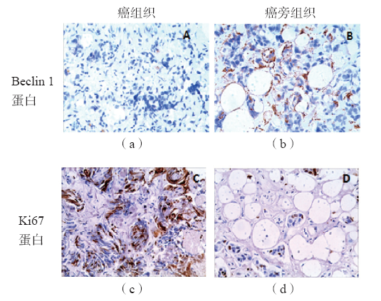
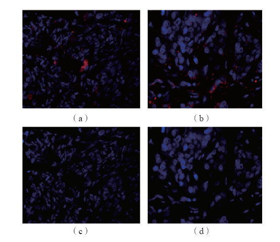
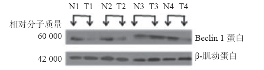
目的 探讨自噬相关基因Beclin 1和增殖相关基因Ki67编码的蛋白在乳腺浸润性导管癌中的表达情况。方法 选取乳腺浸润性导管癌患者64例。采用免疫组化法检测癌组织及对应的癌旁组织中Beclin 1和Ki67蛋白的表达,同时采用免疫荧光法和免疫印迹法检测癌组织及对应癌旁组织中Beclin 1蛋白的表达。结果 癌组织中Beclin 1的阳性表达率(43.8%)明显低于癌旁组织(98.5%)(P<0.05),而Ki67的阳性表达率(76.6%)则高于癌旁组织(0.00%)(P<0.05)。癌组织中Beclin 1蛋白表达与Ki67蛋白表达呈负相关(r=-0.256,P<0.05)。Beclin 1蛋白表达与乳腺浸润性导管癌病理分级、有无淋巴结转移有关(P<0.05),与肿瘤直径、年龄、雌激素受体(ER)、孕激素受体(PR)、人类表皮生长因子受体2(HER-2)均无关(P>0.05)。Ki67蛋白表达与乳腺浸润性导管癌病理分级、肿瘤直径有关(P<0.05),与有无淋巴结转移、年龄、ER、PR、HER-2均无关(P>0.05)。免疫荧光法检测结果显示癌组织中Beclin 1蛋白的阳性表达率(30.0%)明显低于癌旁组织(100.0%)(P<0.05)。免疫印迹法检测结果显示癌旁组织中Beclin l蛋白的相对表达量为0.43±0.12,明显高于癌组织(0.20±0.18)(P<0.05)。结论乳腺浸润性导管癌组织中Beclin 1蛋白表达明显降低,Ki67蛋白表达明显升高。

关键词: Beclin 1基因, Ki67基因, 细胞自噬, 细胞增殖, 乳腺浸润性导管癌

Abstract:

Objective To investigate the expression of autophagy-associated gene Beclin 1 and proliferation-related gene Ki67 in breast invasive ductal carcinoma. Methods The expressions of Beclin 1 and Ki67 were determined by immunohistochemistry in 64 patients with breast invasive ductal carcinoma and corresponding adjacent tissues. Immunofluorescence and western blotting were used to determine the expressions of Beclin 1 in breast invasive ductal carcinoma and corresponding adjacent tissues. Results The positive expression rate of Beclin 1 in carcinoma tissues(43.8%) was lower than that in adjacent tissues(98.5%)(P<0.05). The positive expression rate of Ki67(76.6%) was higher than that in adjacent tissues(0.00%)(P<0.05). The positive rates of Beclin 1 and Ki67 in carcinoma tissues were negatively correlated(r=-0.256,P<0.05). The expression of Beclin 1 was associated with pathological grade and lymph node metastasis(P<0.05),but it was not associated with tumor diameter,age,estrogen receptor(ER),progesterone receptor(PR) and human epidermal growth factor receptor 2(HER-2)(P>0.05). The expression of Ki67 was associated with pathological grade and tumor diameter(P<0.05),but it was not associated with lymph node metastasis,age,ER,PR and HER-2(P>0.05). The results of immunofluorescence showed that the positive expression rate of Beclin 1 in carcinoma tissues(30.0%) was lower than that in adjacent tissues(100.0%)(P<0.05). The results of western blotting showed that the relative expression of Beclin 1 in adjacent tissues was 0.43±0.12,which was higher than that in carcinoma tissues(0.20±0.18)(P<0.05). Conclusions The expression of Beclin 1 is down-regulated in breast invasive ductal carcinoma,while the expression of Ki67 is up-regulated .

Key words: Beclin 1, Ki67, Cell autophagy, Cell proliferation, Breast invasive ductal carcinoma

中图分类号: