检验医学 ›› 2018, Vol. 33 ›› Issue (6): 472-475.DOI: 10.3969/j.issn.1673-8640.2018.06.002

• 临床应用研究?论著 • 上一篇    下一篇

不育患者凋亡生精细胞与ACP超微结构的关系

王平1, 戴研平2(), 杨薇3, 岳应权1, 高晓勤1   

  1. 1. 遵义医药高等专科学校,贵州 遵义 563002
    2. 贵州医科大学基础医学院,贵州 贵阳 550004
    3. 贵阳护理职业学院检验系,贵州 贵阳 550004
  • 收稿日期:2017-04-22 出版日期:2018-06-30 发布日期:2018-07-06
  • 作者简介:null

    作者简介:王平,男,1963年生,副教授,主要从事男性生殖学研究。

  • 基金资助:
    贵州省科学技术基金项目(黔科合人才团队[2014]4005号);遵义市科技计划(遵市科合设字[2014]01号);贵州省科学技术基金项目(黔科合人才团队[2015]7568号)

Relationship of the ultrastructure of acid phosphatase and the apoptosis of spermatogenic cells in infertile patients

WANG Ping1, DAI Yanping2(), YANG Wei3, YUE Yingquan1, GAO Xiaoqin1   

  1. 1. Zunyi Medical and Pharmaceutical College,Zunyi 563002,Guizhou,China
    2. Department of Basic Medicine,Guizhou Medical University,Guiyang 550004,Guizhou,China
    3. Faculty of Laboratory Medicine,Guiyang Nursing Vocational College,Guiyang 550004,Guizhou,China
  • Received:2017-04-22 Online:2018-06-30 Published:2018-07-06

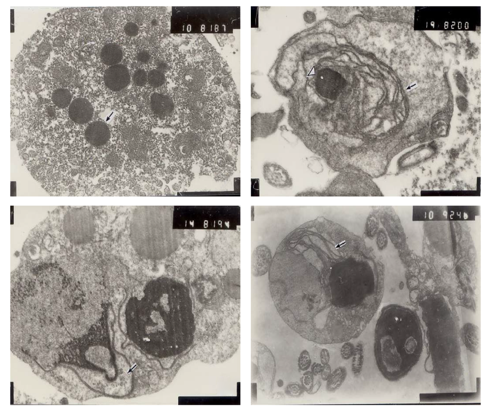
摘要:

目的 研究超微结构下不育患者凋亡生精细胞与酸性磷酸酶(ACP)之间的关系,为男性不育的诊断和治疗提供实验依据。方法 提取正常生育组和不育组男性精液标本各10份,经洗涤、离心和固定后用硫酸铅法处理标本,常规孵育,树脂包埋,在超薄切片后用酶组化电镜观察凋亡生精细胞与ACP超微结构的变化。结果 酶组化电镜观察结果显示,正常生育组精子形态结构基本正常,顶体、质膜、线粒体结构完整,染色质形态规则;不育组可见较多未成熟的生精细胞、巨噬细胞和中性粒细胞内大量与铅结合的ACP沉淀于溶酶体;凋亡精子顶体多形态异常,可见巨噬细胞吞噬凋亡细胞后形成的髓鞘样结构;凋亡生精细胞核染色质浓缩、电子密度高,核膜膨胀、破裂、解体、间隙增宽,线粒体移位、脱落、膨胀,嵴退化或消失,可见凋亡小体、残余体等。与正常生育组比较,不育组ACP阳性反应率明显升高(P<0.05)。结论 ACP活性、数量与生精细胞凋亡数量密切相关,且生精细胞的异常凋亡可导致男性不育,酶组化电镜的应用有望成为男性不育症诊治的新手段之一。

关键词: 生精细胞, 凋亡, 酸性磷酸酶, 超微结构, 男性不育

Abstract:

Objective To study the relationship of the ultrastructure of acid phosphatase(ACP) and the apoptosis of spermatogenic cells in infertile patients,and to provide a reference for the diagnosis and treatment of male infertility. Methods Semen specimens were collected from 10 fertile males and 10 infertile male patients,and the semen specimens were washed,centrifugalized,hatched with Gromori's method and embedded in epoxy resin. The ultra-thin sections were made,and the changes of the ultrastructure of ACP and spermatogenic cell apoptosis were determined by histochemistry using enzyme electron microscope. Results The results of histochemistry using enzyme electron microscope showed that the sperm morphology was normal in fertility group,the structures of acrosome,plasma membrane and mitochondria were integrity,and chromatin was regularly configurated. There were immature spermatogenic cells,macrophage cells and neutrophil cells in infertility group,lead-contacted ACP were showed in lysosome,and various abnormal types of acrosome in apoptotic spermatozoa were observed. The apoptotic bodies formed when apoptotic spermatozoa were phagocytosed,and myelin-like patterns formed as well. The concentration of chromatin and electron density in the nucleus of apoptotic spermatogenic cells were high,the nuclear membrane were expanded,ruptured and disintegrated,gap became wider,mitochondria crest were degraded,falled off or disappeared,and the form of apoptotic bodies and the cytoplasmic residual bodies were found. Compared with fertility group,the ACP positive reaction rate of infertility group was higher with statistical significance(P<0.05). Conclusions The number of apoptotic spermatogenic cells is related closely to the activity and number of ACP. Apoptotic spermatogenic cells are abnormal for infertile males. Histochemistry using enzyme electron microscope is expected to become one way on diagnosing and treating male infertility.

Key words: Spermatogenic cells, Apoptosis, Acid phosphatase, Ultrastructure, Male infertility

中图分类号: