
Laboratory Medicine ›› 2019, Vol. 34 ›› Issue (2): 116-121.DOI: 10.3969/j.issn.1673-8640.2019.02.005
Previous Articles Next Articles
ZHANG Boke1, HUANG Yi2, LIU Yiwen1, HE Yiqing1, YANG Cuixia1, DU Yan1, ZHANG Guoliang1, GAO Feng2( )
)
Received:2018-08-31
Online:2019-02-28
Published:2019-02-28
CLC Number:
ZHANG Boke, HUANG Yi, LIU Yiwen, HE Yiqing, YANG Cuixia, DU Yan, ZHANG Guoliang, GAO Feng. Combined determination of plasma hyaluronan and the ratio of M2 monocyte/M1 monocyte ratio in breast cancer[J]. Laboratory Medicine, 2019, 34(2): 116-121.
Add to citation manager EndNote|Ris|BibTeX
URL: https://www.shjyyx.com/EN/10.3969/j.issn.1673-8640.2019.02.005
| 组别 | 例数 | 年龄(岁) | 白细胞(×109/L) | 单核细胞(×109/L) | HA(ng/mL) |
|---|---|---|---|---|---|
| 乳腺癌组 | 98 | 50.50±10.19 | 5.79±1.65 | 0.35±0.10 | 71.25(55.79~96.08) |
| 乳腺良性疾病组 | 37 | 49.35±8.26 | 5.86±1.18 | 0.36±0.13 | 49.36(43.83~56.13)* |
| 正常对照组 | 41 | 49.52±9.78 | 5.28±1.22 | 0.32±0.11 | 48.70(45.82~54.64)* |
| 组别 | CA15-3(U/mL) | M1型单核细胞比例(%) | M2型单核细胞比例(%) | M2/M1比值 | |
| 乳腺癌组 | 11.06(7.82~16.30) | 30.85±5.52# | 5.71±1.52 | 0.193±0.070 | |
| 乳腺良性疾病组 | 8.15(6.64~11.82)* | 39.11±4.97 | 3.90±0.98* | 0.087±0.028* | |
| 正常对照组 | 9.82(6.75~12.09)* | 32.58±5.31# | 3.76±1.22* | 0.103±0.035* | |
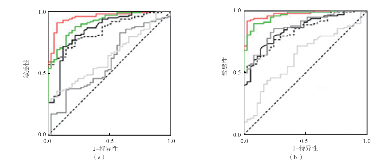
| 项目 | AUC(95%可信区间) | 最佳临界值 | 敏感性(%) | 特异性(%) |
|---|---|---|---|---|
| HA | 0.849(0.787~0.911) | 61.15 ng/mL | 61.22 | 95.12 |
| CA15-3 | 0.638(0.544~0.732) | 13.67 U/mL | 35.71 | 92.68 |
| M1型单核细胞比例 | 0.599(0.495~0.703) | 35.35% | 82.65 | 41.46 |
| M2型单核细胞比例 | 0.852(0.779~0.923) | 4.99% | 71.43 | 87.80 |
| M2/M1比值 | 0.909(0.860~0.957) | 0.138 | 80.60 | 85.37 |
| HA+ M2/M1比值联合检测 | 0.952(0.918~0.987) | 90.82 | 92.68 |
| 项目 | AUC(95%可信区间) | 最佳临界值 | 敏感性(%) | 特异性(%) |
|---|---|---|---|---|
| HA | 0.839(0.772~0.905) | 61.17 ng/mL | 60.20 | 89.19 |
| CA15-3 | 0.631(0.529~0.733) | 13.70 U/mL | 34.68 | 83.78 |
| M1型单核细胞比例 | 0.870(0.808~0.931) | 35.69% | 85.71 | 75.68 |
| M2型单核细胞比例 | 0.851(0.785~0.918) | 5.00% | 69.39 | 86.49 |
| M2/M1比值 | 0.960(0.911~0.984) | 0.127 | 89.80 | 91.89 |
| HA+ M2/M1比值联合检测 | 0.973(0.949~0.997) | 91.84 | 97.30 |
| 临床病理特征 | 例数 | CA15-3(U/mL) | M2/M1比值 | HA(ng/mL) |
|---|---|---|---|---|
| 年龄(岁) | ||||
| ≤50 | 46 | 11.25 (8.21~15.62) | 0.186±0.060 | 65.32 (62.15~89.57) |
| >50 | 52 | 10.92 (8.18~15.72) | 0.198±0.070 | 77.15 (68.56~96.28) |
| P值 | 0.822 | 0.368 | 0.176 | |
| 肿瘤大小(cm) | ||||
| ≤2.0 | 39 | 10.58 (7.78~15.75) | 0.183±0.080 | 75.13 (63.52~90.36) |
| >2.0 | 59 | 11.16 (8.26~16.39) | 0.199±0.060 | 81.56 (60.75~91.39) |
| P值 | 0.119 | 0.261 | 0.611 | |
| TNM分期 | ||||
| 0~Ⅰ期 | 32 | 10.53(7.27~12.59) | 0.139±0.050 | 67.98(63.51~82.65) |
| Ⅱ期 | 50 | 12.22(8.39~18.35) | 0.195±0.070 | 72.55(61.37~95.33) |
| Ⅲ~Ⅳ期 | 16 | 12.53(7.63~18.86) | 0.296±0.060 | 80.07(61.21~138.9) |
| P值 | 0.075 | 0.000 | 0.553 | |
| 临床病理特征 | 例数 | CA15-3(U/mL) | M2/M1比值 | HA(ng/mL) |
| 组织学类型 | ||||
| 浸润性导管癌 | 73 | 11.35 (8.21~17.39) | 0.195±0.080 | 71.66 (63.25~90.36) |
| 其他 | 25 | 10.56 (8.52~15.06) | 0.188±0.070 | 74.19 (60.59~102.93) |
| P值 | 0.229 | 0.698 | 0.522 | |
| 组织学分化 | ||||
| 中~高分化 | 36 | 11.16 (8.30~16.66) | 0.151±0.050 | 66.43 (60.75~85.71) |
| 低分化 | 62 | 10.25 (8.10~15.23) | 0.218±0.060 | 82.89 (66.12~112.8) |
| P值 | 0.513 | 0.000 | 0.005 | |
| 淋巴结转移 | ||||
| N0 | 45 | 10.76 (8.15~14.85) | 0.149±0.050 | 71.33 (63.25~89.27) |
| N1~3 | 53 | 12.25 (8.33~17.20) | 0.230±0.070 | 73.36 (61.27~97.29) |
| P值 | 0.208 | 0.000 | 0.611 | |
| ER | ||||
| 阳性 | 68 | 11.17 (8.56~16.07) | 0.165±0.050 | 68.38 (62.15~85.27) |
| 阴性 | 30 | 10.56 (7.88~15.59) | 0.256±0.070 | 81.18 (61.21~119.9) |
| P值 | 0.561 | 0.000 | 0.191 | |
| PR | ||||
| 阳性 | 61 | 10.58 (8.63~15.17) | 0.188±0.070 | 71.32 (61.35~87.59) |
| 阴性 | 37 | 12.15 (7.90~18.19) | 0.201±0.060 | 75.73 (62.28~105.37) |
| P值 | 0.198 | 0.350 | 0.501 | |
| HER-2 | ||||
| 阳性 | 21 | 11.15 (7.12~18.16) | 0.211±0.070 | 65.28 (60.72~85.93) |
| 阴性 | 77 | 10.88 (8.18~15.04) | 0.188±0.060 | 75.08 (63.55~96.17) |
| P值 | 0.794 | 0.137 | 0.102 |
| 临床病理特征 | 例数 | CA15-3(U/mL) | M2/M1比值 | HA(ng/mL) |
|---|---|---|---|---|
| 年龄(岁) | ||||
| ≤50 | 46 | 11.25 (8.21~15.62) | 0.186±0.060 | 65.32 (62.15~89.57) |
| >50 | 52 | 10.92 (8.18~15.72) | 0.198±0.070 | 77.15 (68.56~96.28) |
| P值 | 0.822 | 0.368 | 0.176 | |
| 肿瘤大小(cm) | ||||
| ≤2.0 | 39 | 10.58 (7.78~15.75) | 0.183±0.080 | 75.13 (63.52~90.36) |
| >2.0 | 59 | 11.16 (8.26~16.39) | 0.199±0.060 | 81.56 (60.75~91.39) |
| P值 | 0.119 | 0.261 | 0.611 | |
| TNM分期 | ||||
| 0~Ⅰ期 | 32 | 10.53(7.27~12.59) | 0.139±0.050 | 67.98(63.51~82.65) |
| Ⅱ期 | 50 | 12.22(8.39~18.35) | 0.195±0.070 | 72.55(61.37~95.33) |
| Ⅲ~Ⅳ期 | 16 | 12.53(7.63~18.86) | 0.296±0.060 | 80.07(61.21~138.9) |
| P值 | 0.075 | 0.000 | 0.553 | |
| 临床病理特征 | 例数 | CA15-3(U/mL) | M2/M1比值 | HA(ng/mL) |
| 组织学类型 | ||||
| 浸润性导管癌 | 73 | 11.35 (8.21~17.39) | 0.195±0.080 | 71.66 (63.25~90.36) |
| 其他 | 25 | 10.56 (8.52~15.06) | 0.188±0.070 | 74.19 (60.59~102.93) |
| P值 | 0.229 | 0.698 | 0.522 | |
| 组织学分化 | ||||
| 中~高分化 | 36 | 11.16 (8.30~16.66) | 0.151±0.050 | 66.43 (60.75~85.71) |
| 低分化 | 62 | 10.25 (8.10~15.23) | 0.218±0.060 | 82.89 (66.12~112.8) |
| P值 | 0.513 | 0.000 | 0.005 | |
| 淋巴结转移 | ||||
| N0 | 45 | 10.76 (8.15~14.85) | 0.149±0.050 | 71.33 (63.25~89.27) |
| N1~3 | 53 | 12.25 (8.33~17.20) | 0.230±0.070 | 73.36 (61.27~97.29) |
| P值 | 0.208 | 0.000 | 0.611 | |
| ER | ||||
| 阳性 | 68 | 11.17 (8.56~16.07) | 0.165±0.050 | 68.38 (62.15~85.27) |
| 阴性 | 30 | 10.56 (7.88~15.59) | 0.256±0.070 | 81.18 (61.21~119.9) |
| P值 | 0.561 | 0.000 | 0.191 | |
| PR | ||||
| 阳性 | 61 | 10.58 (8.63~15.17) | 0.188±0.070 | 71.32 (61.35~87.59) |
| 阴性 | 37 | 12.15 (7.90~18.19) | 0.201±0.060 | 75.73 (62.28~105.37) |
| P值 | 0.198 | 0.350 | 0.501 | |
| HER-2 | ||||
| 阳性 | 21 | 11.15 (7.12~18.16) | 0.211±0.070 | 65.28 (60.72~85.93) |
| 阴性 | 77 | 10.88 (8.18~15.04) | 0.188±0.060 | 75.08 (63.55~96.17) |
| P值 | 0.794 | 0.137 | 0.102 |
| [1] | DUFFY M J,EVOY D,MCDERMOTT E W.CA 15-3:uses and limitation as a biomarker for breast cancer[J]. Clin Chim Acta,2010,411(23-24):1869-1874. |
| [2] | SOUSA S,BRION R,LINTUNEN M,et al.Human breast cancer cells educate macrophages toward the M2 activation status[J]. Breast Cancer Res,2015,17:101. |
| [3] | ROUSSOS E T,BALSAMO M,ALFORD S K,et al.Mena invasive (MenaINV) promotes multicellular streaming motility and transendothelial migration in a mouse model of breast cancer[J]. J Cell Sci,2011,124(Pt 13):2120-2131. |
| [4] | ADAMS D L,MARTIN S S,ALPAUGH R K,et al.Circulating giant macrophages as a potential biomarker of solid tumors[J]. Proc Natl Acad Sci U S A,2014,111(9):3514-3519. |
| [5] | SICA A,SCHIOPPA T,MANTOVANI A,et al.Tumour-associated macrophages are a distinct M2 polarised population promoting tumour progression:potential targets of anti-cancer therapy[J]. Eur J Cancer,2006,42(6):717-727. |
| [6] | ZHANG B,CAO M,HE Y,et al.Increased circulating M2-like monocytes in patients with breast cancer[J]. Tumour Biol,2017,39(6):1010428317711571. |
| [7] | WU M,CAO M,HE Y,et al.A novel role of low molecular weight hyaluronan in breast cancer metastasis[J]. FASEB J,2015,29(4):1290-1298. |
| [8] | TIAINEN S,TUMELIUS R,RILLA K,et al.High numbers of macrophages,especially M2-like(CD163-positive),correlate with hyaluronan accumulation and poor outcome in breast cancer[J]. Histopathology,2015,66(6):873-883. |
| [9] | MANTOVANI A,SOZZANI S,LOCATI M,et al.Macrophage polarization: tumor-associated macrophages as a paradigm for polarized M2 mononuclear phagocytes[J]. Trends Immunol,2002,23(11):549-555. |
| [10] | MAEDA R,ISHII G,NERI S,et al.Circulating CD14+CD204+ cells predict postoperative recurrence in non-small-cell lung cancer patients[J]. J Thorac Oncol,2014,9(2):179-188. |
| [11] | LI C,LUO X,LIN Y,et al.A higher frequency of CD14+ CD169+ monocytes/macrophages in patients with colorectal cancer[J]. PLoS One,2015,10(10):e0141817. |
| [12] | OSTUNI R,KRATOCHVILL F,MURRAY P J,et al.Macrophages and cancer:from mechanisms to therapeutic implications[J]. Trends Immunol,2015,36(4):229-239. |
| [13] | CLAWSON G A,MATTERS G L,XIN P,et al.Macrophage-tumor cell fusions from peripheral blood of melanoma patients[J]. PLoS One,2015,10(8):e0134320. |
| [14] | KUANG D M,WU Y,CHEN N,et al.Tumor-derived hyaluronan induces formation of immunosuppressive macrophages through transient early activation of monocytes[J]. Blood,2007,110(2):587-595. |
| [1] | HUANG Lei, TANG Wenjia, ZHOU Yan, ZHOU Jiaye, ZHANG Chunyan, YANG Jing, WANG Beili, PAN Baishen, GUO wei. Prognosis of RDW-SD and RDW-CV in breast cancer patients with lymphatic metastasis [J]. Laboratory Medicine, 2024, 39(4): 376-381. |
| [2] | CUI Xiaoyang, LIU Guodong, GUO Huijuan, LIAO Zhihong, LIU Yunhong, ZHANG Weifen, CHEN Zirao, WEI Xiaozhu. Roles of serum exosomal CEA,CA15-3 and CA125 in the differential diagnosis of non-small cell lung cancer and benign lung diseases [J]. Laboratory Medicine, 2024, 39(11): 1035-1041. |
| [3] | LIU Bohan, LIU Yiwen, HE Yiqing, LU Renquan, DU Yan, ZHANG Guoliang, GUO Qian, GAO Feng, YANG Cuixia. Serum amphiregulin and mesothelin in auxiliary diagnosis of breast cancer [J]. Laboratory Medicine, 2024, 39(1): 26-30. |
| [4] | WANG Rong, XING Lianxiang, HUANG Keliang, LI Xin. MiR-374 promoting proliferation and invasion of breast cancer cells by targeting and down-regulating TRIM35 [J]. Laboratory Medicine, 2023, 38(9): 812-817. |
| [5] | CHEN Chen, DUAN Qi, LU Jiatuan, ZHAI Xiaojian, WANG Zheng, ZHANG Hao, GUO Man. Expressions of ASH2L and HOXA2 in triple-negative breast cancer patients and their relationship with lymph node metastasis [J]. Laboratory Medicine, 2023, 38(6): 574-578. |
| [6] | ZHANG Guoliang, LIU Yiwen, HE Yiqing, XU Jing, YANG Cuixia, GAO Feng, LIU Hua. Changes of serum HAS2 and CD44 levels in breast cancer patients and their clinical significance [J]. Laboratory Medicine, 2023, 38(5): 424-429. |
| [7] | ZHANG Guoliang, LIU Yiwen, HE Yiqing, XU Jing, YANG Cuixia, GAO Feng, LIU Hua. Roles of serum HYAL1 and HA in the auxiliary diagnosis and therapeutic monitoring of colorectal cancer [J]. Laboratory Medicine, 2023, 38(4): 313-319. |
| [8] | LIU Qinqing, XU Jing, LIU Hua, LIU Yiwen, HE Yiqing, DU Yan, ZHANG Guoliang, GAO Feng, YANG Cuixia. Role of hyaluronan in thyroid nodule fine-needle aspiration washout fluid in differential diagnosis of papillary thyroid cancer [J]. Laboratory Medicine, 2023, 38(4): 320-324. |
| [9] | LI Mu, GONG Dongliang, XU Liming, PENG Rong. Relationship of XPC rs2228000 polymorphisms and breast cancer [J]. Laboratory Medicine, 2023, 38(3): 235-239. |
| [10] | LIU Chong, ZHANG Jing, LI Sheng, ZHAO Qi. Expressions and correlation of miR-335 and Fra-1 in breast cancer [J]. Laboratory Medicine, 2023, 38(2): 143-147. |
| [11] | GAO Jianchao, WANG Sisi, ZHANG Zhisheng, ZHANG Jingli, LI Xiaoxia, MA Ke, FENG Zhilin, ZHOU Haifeng, WANG Zhanhai. Efficacy and prognosis evaluation of neo-adjuvant chemotherapy for breast cancer based on miR-206,miR-125 and miR-21 [J]. Laboratory Medicine, 2023, 38(11): 1062-1068. |
| [12] | WANG Xiaoye, DONG Guoyou, LIU Zhiying. Relations of ZEB2 and E-Cad expressions in breast cancer tissues with prognosis [J]. Laboratory Medicine, 2022, 37(9): 815-820. |
| [13] | YU Fangfang, ZHAO Qi, YANG Liping, WANG Chenyu. Correlation of hematological indexes and expression of HER-2 in patients with breast cancer [J]. Laboratory Medicine, 2022, 37(6): 514-517. |
| [14] | YE Jingwen, SHEN Yunyue, LIU Yiwen, HE Yiqing, DU Yan, ZHANG Guoliang, GAO Feng, YANG Cuixia. Role of MAPK/ERK signaling pathway in reversing endocrine resistance of breast cancer [J]. Laboratory Medicine, 2022, 37(4): 342-348. |
| [15] | ZHANG Xinyue, CHEN Liang, ZHENG Yu. Correlation between serum C peptide and insulin-like growth factor binding protein 3 and the risk of breast cancer patient death [J]. Laboratory Medicine, 2022, 37(1): 36-40. |
| Viewed | ||||||
|
Full text |
|
|||||
|
Abstract |
|
|||||